Showing posts with label breast cancer detection. Show all posts

Wednesday, March 24, 2021

Abstract-Terahertz refractive index-based morphological dilation for breast carcinoma delineation


Quentin Cassar, Samuel Caravera, Gaëtan MacGrogan, Thomas Bücher, Philipp Hillger, Ullrich Pfeiffer, Thomas Zimmer, Jean-Paul Guillet,  Patrick Mounaix


https://www.nature.com/articles/s41598-021-85853-8

This paper reports investigations led on the combination of the refractive index and morphological dilation to enhance performances towards breast tumour margin delineation during conserving surgeries. The refractive index map of invasive ductal and lobular carcinomas were constructed from an inverse electromagnetic problem. Morphological dilation combined with refractive index thresholding was conducted to classify the tissue regions as malignant or benign. A histology routine was conducted to evaluate the performances of various dilation geometries associated with different thresholds. It was found that the combination of a wide structuring element and high refractive index was improving the correctness of tissue classification in comparison to other configurations or without dilation. The method reports a sensitivity of around 80% and a specificity of 82% for the best case. These results indicate that combining the fundamental optical properties of tissues denoted by their refractive index with morphological dilation may open routes to define supporting procedures during breast-conserving surgeries.

Tuesday, October 27, 2020

Terahertz Waves Can Image Early-Stage Breast Cancer Without Staining


https://www.technologynetworks.com/tn/news/terahertz-waves-can-image-early-stage-breast-cancer-without-staining-341852

A team of researchers at Osaka University, in collaboration with the University of Bordeaux and the Bergonié Institute in France, has succeeded in terahertz imaging of early-stage breast cancer less than 0.5 mm without staining, which is difficult to identify even by pathological diagnosis. Their work provides a breakthrough towards rapid and precise on-site diagnosis of various types of cancer and accelerates the development of innovative terahertz diagnostic devices.


Breast cancer is roughly divided into two types: invasive and non-invasive. The former, invasive ductal carcinoma (IDC), begins in the cells of a breast duct, growing through the duct walls and into the surrounding breast tissue, potentially spreading to other parts of the body. The latter, ductal carcinoma in situ (DCIS), is an early-stage small breast cancer confined to the breast duct, but it can lead to invasive cancer. Therefore, early detection of DCIS is crucial.

For pathological diagnosis of cancer, the tissue sample is chemically stained, and a pathologist makes a diagnosis using an image of the stained tissue. However, the staining process takes time, and it is difficult to distinguish DCIS from malignant IDC as they look nearly identical.

Terahertz imaging can distinguish cancer tissue from normal tissue without staining and radiation exposure. However, it was still difficult to identify an individual DCIS lesion (which typically range from 50 to 500 μm) by terahertz imaging due to its diffraction-limited spatial resolution of just several millimeters.

"To overcome this drawback, we developed a unique imaging technique in which terahertz light sources that are locally generated at irradiation spots of laser beams in a nonlinear optical crystal directly interact with a breast cancer tissue sample. Consequently, we succeeded in clearly visualizing a DCIS lesion of less than 0.5 mm," explains lead author Kosuke Okada. The accuracy of this technique is approximately 1000 times higher than that of conventional techniques using terahertz waves.

The researchers also found that terahertz intensity distributions were different between DCIS and IDC, suggesting the possibility of quantitative determination of cancer malignancy.

The breast cancer tissue sample was provided and histologically assessed by collaborators from the University of Bordeaux and the Bergonié Institute. "One of the challenges in this research is preparing a high-quality breast cancer tissue sample fabricated on a nonlinear optical crystal. It is one of the great achievements of international joint research," says corresponding author Masayoshi Tonouchi.

"Combining our technique with machine learning will aid in the early detection of cancer and determination of cancer malignancy, as well as development of innovative terahertz diagnostic devices using Micro Electro Mechanical Systems."


Reference: The article, “Terahertz near-field microscopy of ductal carcinoma in situ (DCIS) of the breast,” will be published online on Oct. 22, 2020 in Journal of Physics: Photonics at DOI: https://doi.org/10.1088/2515-7647/abbcda.

Friday, May 24, 2019

Abstract-Terahertz tomographic imaging of freshly excised human breast tissues


Tyler Bowman,  Nagma Vohra,  Keith Bailey, Magda O. El-Shenawee

https://www.spiedigitallibrary.org/journals/Journal-of-Medical-Imaging/volume-6/issue-2/023501/Terahertz-tomographic-imaging-of-freshly-excised-human-breast-tissues/10.1117/1.JMI.6.2.023501.short?SSO=1


Terahertz imaging and spectroscopy characterization of freshly excised breast cancer tumors are presented in the range 0.15 to 3.5 THz. Cancerous breast tissues were obtained from partial or full removal of malignant tumors while healthy breast tissues were obtained from breast reduction surgeries. The reflection spectroscopy to obtain the refractive index and absorption coefficient is performed on experimental data at each pixel of the tissue, forming tomographic images. The transmission spectroscopy of the refractive index and absorption coefficient are retrieved from experimental data at few tissue points. The average refractive index and absorption coefficients for cancer, fat, and collagen tissue regions are compared between transmission and reflection modes. The reflection mode offers the advantage of retrieving the electrical properties across a significantly greater number of points without the need for sectioning or altering the freshly excised tissue as in the transmission mode. The terahertz spectral power images and the tomographic images demonstrated good qualitative comparison with pathology.
© 2019 Society of Photo-Optical Instrumentation Engineers (SPIE) 2329-4302/2019/$25.00 © 2019 SPIE

Tuesday, April 30, 2019

Abstract-High-sensitivity detection of metastatic breast cancer cells via terahertz chemical microscopy using aptamers


Eman M.Hassan, Ahmed Mohamed, Maria C.DeRosa, William G.Willmore, Yuki Hanaoka, Toshihiko Kiwa, Tsuneyuki Ozaki

Fig. 1. Schematic diagram of the optical setup of the TCM used in this study
https://www.sciencedirect.com/science/article/pii/S0925400519302229?via%3Dihub

We demonstrate high-sensitivity detection of metastatic breast cancer cells using terahertz (THz) chemical microscopy (TCM) with aptamers as ligands. For the aptamers, we use previously developed synthetic single stranded (ss) DNA aptamers mammaglobin B1 (MAMB1) and mammaglobin A2 (MAMA2) that bind to mammaglobin B and mammaglobin A proteins, respectively, which are overexpressed on the surface of MCF7 and MDA-MB-415 breast cancer cells. Each aptamer was immobilized on the surface of a sensing plate, and the amplitude of the THz signal was measured upon the binding of each aptamer to different number (10–106) of its target breast cancer cells. A change in the THz amplitude was observed when MAMB1 and MAMA2 bind to MCF7 and MDA-MB-415, respectively. We find that this change was linear as a function of the log number of breast cancer cells used. No change in the THz amplitude was observed when the same number of normal breast cells (MCF 10A) were used. Moreover, MAMB1 and MAMA2 did not show binding to the counter breast cancer cells, indicating high selectivity. We have demonstrated that the TCM using aptamers as ligands has a limit of detection as small as 1 breast cancer cell in 100 μL of sample. Results described here indicate that the TCM could be a powerful tool to detect metastatic breast cancer cells.

Friday, August 10, 2018

Abstract-Assessment of Terahertz Imaging for Excised Breast Cancer Tumors with Image Morphing


Tanny Chavez, Tyler Bowman, Jingxian Wu, Keith Bailey, Magda El-Shenawee

https://link.springer.com/article/10.1007/s10762-018-0529-8


This paper presents an image morphing algorithm for quantitative evaluation methodology of terahertz (THz) images of excised breast cancer tumors. Most current studies on the assessment of THz imaging rely on qualitative evaluation, and there is no established benchmark or procedure to quantify the THz imaging performance. The proposed morphing algorithm provides a tool to quantitatively align the THz image with the histopathology image. Freshly excised xenograft murine breast cancer tumors are imaged using the pulsed THz imaging and spectroscopy system in the reflection mode. Upon fixing the tumor tissue in formalin and embedding in paraffin, a formalin-fixed paraffin-embedded (FFPE) tissue block is produced. A thin slice of the block is prepared for the pathology image while another THz reflection image is produced directly from the block. We developed an algorithm of mesh morphing using homography mapping of the histopathology image to adjust the alignment, shape, and resolution to match the external contour of the tissue in the THz image. Unlike conventional image morphing algorithms that rely on internal features of the source and target images, only the external contour of the tissue is used to avoid bias. Unsupervised Bayesian learning algorithm is applied to THz images to classify the tissue regions of cancer, fat, and muscles present in xenograft breast tumors. The results demonstrate that the proposed mesh morphing algorithm can provide more effective and accurate evaluation of THz imaging compared with existing algorithms. The results also showed that while THz images of FFPE tissue are highly in agreement with pathology images, challenges remain in assessing THz imaging of fresh tissue.

Thursday, March 1, 2018

Researchers Move Closer to Improved Method of Detecting Breast Cancer


                                                           Magda El-Shenawee

https://news.uark.edu/articles/41165/researchers-move-closer-to-improved-method-of-detecting-breast-cancer

FAYETTEVILLE, Ark. – Engineering researchers at the University of Arkansas have moved closer to developing an alternative method of detecting and possibly treating breast cancer.
The researchers, led by Magda El-Shenawee, professor of electrical engineering, work with pulsed, terahertz imaging, a type of electromagnetic radiation technology previously used to find land mines. They adapted the technology to detect tumors and provide highly specific images of them.
Standard breast cancer imaging techniques do not always provide clear assessment of breast tissue on the margins of a tumor. Without an accurate picture of the margins between the tumor and healthy tissue, surgeons cannot be sure they have removed the entire tumor during a surgery.
This shortcoming contributes to high rates – 20 to 40 percent – of secondary surgery, either lumpectomy or mastectomy. Terahertz imaging could lead to fast, noninvasive, and highly specific tumor margin assessment, which in turn could reduce the occurrence of second surgeries, cancer reoccurrence and metastasis.

FINDINGS

In their most recent study, funded by a $424,081 grant from the National Institutes of Health, the researchers created terahertz images of breast adenocarcinoma cells, a type of malignancy, excised from mice. These images were taken from 13 tumor samples. To test the accuracy of the terahertz method, the images were then statistically compared to high-resolution histopathology images of the same excised tumor samples. Histopathology is the microscopic examination of tissue changes caused by disease.
In all tissue samples containing only cancer and fat – a combination similar to tissue in the human breast – the terahertz images, when compared to the histopathology images, accurately detected cancerous tissue with a high level of specificity.
On the other hand, samples containing cancer, fat and muscle showed only a reasonable correlation, El-Shenawee said. In these samples, the terahertz images detected cancer cells at the margins of tumors and muscle tissue, but not at high enough levels of specificity.
“Overlap between muscle and cancer tissue in the terahertz image creates some challenge in correctly classifying these regions,” El Shenawee said. “While muscle is unlikely to be present in surgical sections of human breast cancer, other kinds of fibrous tissue may be, so this requires further investigation with more advanced models.”
Future work will focus on spontaneously generated breast cancer tumors from genetically modified mice, which have tumor and tissue structures closer to that of humans. El-Shenawee said these models will provide a more accurate assessment of terahertz imaging. The researchers will also compare the terahertz images to other standard imaging techniques, such as radiography and computed tomography, or CT scan.

TERAHERTZ IMAGING
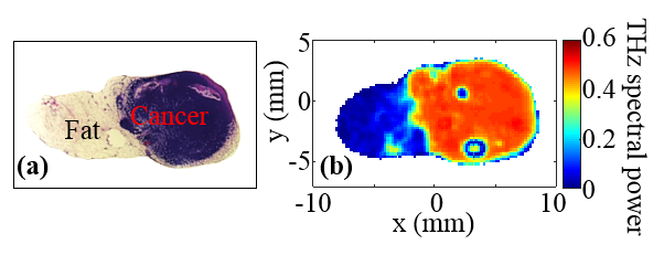

Pathology image, left, and corresponding terahertz image, right, of excised tissue from mouse breast tumor. Photo submitted by the researcher.
Pulsed, terahertz spectroscopy produces high-quality images of the tissue, down to 80 micrometers. It scatters fewer waves than radiography, which enables deeper imaging into an object. Also, because terahertz radiation can transmit through most non-metallic materials, the systems can “see” through concealing barriers. For many years, El-Shenawee has focused on developing this detection system for health-care applications, while also investigating the unique electromagnetic signals emitted by breast cancer cells.

THE TEAM

The research team included Tyler Bowman, Tanny Chavez, graduate students in electrical engineering; Jingxian Wu, associate professor of electrical engineering; Kamrul Khan, graduate student in mathematical sciences; Avishek Chakraborty, assistant professor of statistics; Narasimhan Rajaram, assistant professor of biomedical engineering; and Keith Bailey, animal pathologist at Oklahoma State University.
Their findings were published in the February issue of Journal of Biomedical Optics.
This research was conducted in the Terahertz Imaging and Spectroscopy research facilities at the University of Arkansas.
About the College of Engineering: The University of Arkansas College of Engineering is the largest engineering program in the state of Arkansas. Over the past decade, the college has experienced unprecedented growth. Undergraduate enrollment has doubled since 2007, and total enrollment in the college is now over 4,000 students. The College of Engineering offers graduate and undergraduate degrees in nine engineering fields, as well as incorporating distance learning and interdisciplinary programs. Faculty in the college conduct research in many key areas, including electronics, energy, biomedical and healthcare engineering, materials science, transportation and logistics. 
About the University of Arkansas: The University of Arkansas provides an internationally competitive education for undergraduate and graduate students in more than 200 academic programs. The university contributes new knowledge, economic development, basic and applied research, and creative activity while also providing service to academic and professional disciplines. The Carnegie Foundation classifies the University of Arkansas among only 2 percent of universities in America that have the highest level of research activity. U.S. News & World Report ranks the University of Arkansas among its top American public research universities. Founded in 1871, the University of Arkansas comprises 10 colleges and schools and maintains a low student-to-faculty ratio that promotes personal attention and close mentoring.
CONTACTS
Magda El-Shenawee, professor, electrical engineering 
College of Engineering 
479-575-6582, magda@uark.edu
Matt McGowan, science and research communications officer 
University Relations 
479-575-4246, dmcgowa@uark.edu