Showing posts with label Tyler Bowman. Show all posts

Friday, June 12, 2020

Abstract-Terahertz Time-Domain Pulsed Spectroscopy of Human Breast Cancer Tissues


Tyler BowmanMagda El-ShenaweeKeith Bailey

breast tumors using terahertz imaging ...
content.iospress.com
https://ieeexplore.ieee.org/document/8608929

This work utilizes time-domain pulsed terahertz spectroscopy to obtain the refractive index and absorption coefficient of normal and malignant human breast cancer tissues. The excised tissues are purchased from the National Disease Research Interchange and shipped within 24 hours of surgery. The tissue is immersed in Dulbecco's Modified Eagle Medium plus antibiotic during the shipment. The solution is characterized in the terahertz frequency band (0.1-4 THz)showing insignificant differences compared with distilled water and other common solutions. Healthy fibrous and fatty breast tissue are obtained from breast reduction surgery, while malignant tumors are obtained from breast cancer mastectomy or lumpectomy surgeries. The results will help understand the interaction of terahertz waves with human breast tumors and also to develop phantom tissue that can be manipulated to advance terahertz imaging.

Friday, May 24, 2019

Abstract-Terahertz tomographic imaging of freshly excised human breast tissues


Tyler Bowman,  Nagma Vohra,  Keith Bailey, Magda O. El-Shenawee

https://www.spiedigitallibrary.org/journals/Journal-of-Medical-Imaging/volume-6/issue-2/023501/Terahertz-tomographic-imaging-of-freshly-excised-human-breast-tissues/10.1117/1.JMI.6.2.023501.short?SSO=1


Terahertz imaging and spectroscopy characterization of freshly excised breast cancer tumors are presented in the range 0.15 to 3.5 THz. Cancerous breast tissues were obtained from partial or full removal of malignant tumors while healthy breast tissues were obtained from breast reduction surgeries. The reflection spectroscopy to obtain the refractive index and absorption coefficient is performed on experimental data at each pixel of the tissue, forming tomographic images. The transmission spectroscopy of the refractive index and absorption coefficient are retrieved from experimental data at few tissue points. The average refractive index and absorption coefficients for cancer, fat, and collagen tissue regions are compared between transmission and reflection modes. The reflection mode offers the advantage of retrieving the electrical properties across a significantly greater number of points without the need for sectioning or altering the freshly excised tissue as in the transmission mode. The terahertz spectral power images and the tomographic images demonstrated good qualitative comparison with pathology.
© 2019 Society of Photo-Optical Instrumentation Engineers (SPIE) 2329-4302/2019/$25.00 © 2019 SPIE

Friday, August 10, 2018

Abstract-Assessment of Terahertz Imaging for Excised Breast Cancer Tumors with Image Morphing


Tanny Chavez, Tyler Bowman, Jingxian Wu, Keith Bailey, Magda El-Shenawee

https://link.springer.com/article/10.1007/s10762-018-0529-8


This paper presents an image morphing algorithm for quantitative evaluation methodology of terahertz (THz) images of excised breast cancer tumors. Most current studies on the assessment of THz imaging rely on qualitative evaluation, and there is no established benchmark or procedure to quantify the THz imaging performance. The proposed morphing algorithm provides a tool to quantitatively align the THz image with the histopathology image. Freshly excised xenograft murine breast cancer tumors are imaged using the pulsed THz imaging and spectroscopy system in the reflection mode. Upon fixing the tumor tissue in formalin and embedding in paraffin, a formalin-fixed paraffin-embedded (FFPE) tissue block is produced. A thin slice of the block is prepared for the pathology image while another THz reflection image is produced directly from the block. We developed an algorithm of mesh morphing using homography mapping of the histopathology image to adjust the alignment, shape, and resolution to match the external contour of the tissue in the THz image. Unlike conventional image morphing algorithms that rely on internal features of the source and target images, only the external contour of the tissue is used to avoid bias. Unsupervised Bayesian learning algorithm is applied to THz images to classify the tissue regions of cancer, fat, and muscles present in xenograft breast tumors. The results demonstrate that the proposed mesh morphing algorithm can provide more effective and accurate evaluation of THz imaging compared with existing algorithms. The results also showed that while THz images of FFPE tissue are highly in agreement with pathology images, challenges remain in assessing THz imaging of fresh tissue.

Thursday, March 1, 2018

Researchers Move Closer to Improved Method of Detecting Breast Cancer


                                                           Magda El-Shenawee

https://news.uark.edu/articles/41165/researchers-move-closer-to-improved-method-of-detecting-breast-cancer

FAYETTEVILLE, Ark. – Engineering researchers at the University of Arkansas have moved closer to developing an alternative method of detecting and possibly treating breast cancer.
The researchers, led by Magda El-Shenawee, professor of electrical engineering, work with pulsed, terahertz imaging, a type of electromagnetic radiation technology previously used to find land mines. They adapted the technology to detect tumors and provide highly specific images of them.
Standard breast cancer imaging techniques do not always provide clear assessment of breast tissue on the margins of a tumor. Without an accurate picture of the margins between the tumor and healthy tissue, surgeons cannot be sure they have removed the entire tumor during a surgery.
This shortcoming contributes to high rates – 20 to 40 percent – of secondary surgery, either lumpectomy or mastectomy. Terahertz imaging could lead to fast, noninvasive, and highly specific tumor margin assessment, which in turn could reduce the occurrence of second surgeries, cancer reoccurrence and metastasis.

FINDINGS

In their most recent study, funded by a $424,081 grant from the National Institutes of Health, the researchers created terahertz images of breast adenocarcinoma cells, a type of malignancy, excised from mice. These images were taken from 13 tumor samples. To test the accuracy of the terahertz method, the images were then statistically compared to high-resolution histopathology images of the same excised tumor samples. Histopathology is the microscopic examination of tissue changes caused by disease.
In all tissue samples containing only cancer and fat – a combination similar to tissue in the human breast – the terahertz images, when compared to the histopathology images, accurately detected cancerous tissue with a high level of specificity.
On the other hand, samples containing cancer, fat and muscle showed only a reasonable correlation, El-Shenawee said. In these samples, the terahertz images detected cancer cells at the margins of tumors and muscle tissue, but not at high enough levels of specificity.
“Overlap between muscle and cancer tissue in the terahertz image creates some challenge in correctly classifying these regions,” El Shenawee said. “While muscle is unlikely to be present in surgical sections of human breast cancer, other kinds of fibrous tissue may be, so this requires further investigation with more advanced models.”
Future work will focus on spontaneously generated breast cancer tumors from genetically modified mice, which have tumor and tissue structures closer to that of humans. El-Shenawee said these models will provide a more accurate assessment of terahertz imaging. The researchers will also compare the terahertz images to other standard imaging techniques, such as radiography and computed tomography, or CT scan.

TERAHERTZ IMAGING
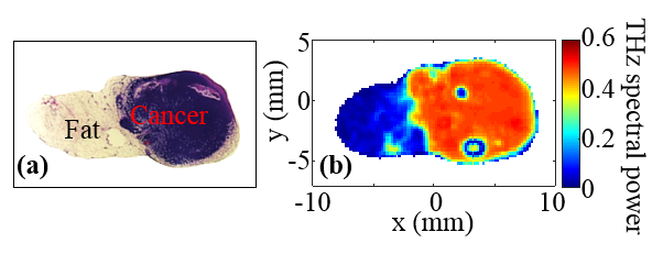

Pathology image, left, and corresponding terahertz image, right, of excised tissue from mouse breast tumor. Photo submitted by the researcher.
Pulsed, terahertz spectroscopy produces high-quality images of the tissue, down to 80 micrometers. It scatters fewer waves than radiography, which enables deeper imaging into an object. Also, because terahertz radiation can transmit through most non-metallic materials, the systems can “see” through concealing barriers. For many years, El-Shenawee has focused on developing this detection system for health-care applications, while also investigating the unique electromagnetic signals emitted by breast cancer cells.

THE TEAM

The research team included Tyler Bowman, Tanny Chavez, graduate students in electrical engineering; Jingxian Wu, associate professor of electrical engineering; Kamrul Khan, graduate student in mathematical sciences; Avishek Chakraborty, assistant professor of statistics; Narasimhan Rajaram, assistant professor of biomedical engineering; and Keith Bailey, animal pathologist at Oklahoma State University.
Their findings were published in the February issue of Journal of Biomedical Optics.
This research was conducted in the Terahertz Imaging and Spectroscopy research facilities at the University of Arkansas.
About the College of Engineering: The University of Arkansas College of Engineering is the largest engineering program in the state of Arkansas. Over the past decade, the college has experienced unprecedented growth. Undergraduate enrollment has doubled since 2007, and total enrollment in the college is now over 4,000 students. The College of Engineering offers graduate and undergraduate degrees in nine engineering fields, as well as incorporating distance learning and interdisciplinary programs. Faculty in the college conduct research in many key areas, including electronics, energy, biomedical and healthcare engineering, materials science, transportation and logistics. 
About the University of Arkansas: The University of Arkansas provides an internationally competitive education for undergraduate and graduate students in more than 200 academic programs. The university contributes new knowledge, economic development, basic and applied research, and creative activity while also providing service to academic and professional disciplines. The Carnegie Foundation classifies the University of Arkansas among only 2 percent of universities in America that have the highest level of research activity. U.S. News & World Report ranks the University of Arkansas among its top American public research universities. Founded in 1871, the University of Arkansas comprises 10 colleges and schools and maintains a low student-to-faculty ratio that promotes personal attention and close mentoring.
CONTACTS
Magda El-Shenawee, professor, electrical engineering 
College of Engineering 
479-575-6582, magda@uark.edu
Matt McGowan, science and research communications officer 
University Relations 
479-575-4246, dmcgowa@uark.edu

Friday, February 23, 2018

Abstract-Pulsed terahertz imaging of breast cancer in freshly excised murine tumors


Tyler Bowman, Tanny Chavez, Kamrul Khan,  Jingxian Wu,  Avishek Chakraborty,  Narasimhan Rajaram, Keith Bailey,  Magda El-Shenawee,


https://www.spiedigitallibrary.org/journals/Journal-of-Biomedical-Optics/volume-23/issue-2/026004/Pulsed-terahertz-imaging-of-breast-cancer-in-freshly-excised-murine/10.1117/1.JBO.23.2.026004.short


This paper investigates terahertz (THz) imaging and classification of freshly excised murine xenograft breast cancer tumors. These tumors are grown via injection of E0771 breast adenocarcinoma cells into the flank of mice maintained on high-fat diet. Within 1 h of excision, the tumor and adjacent tissues are imaged using a pulsed THz system in the reflection mode. The THz images are classified using a statistical Bayesian mixture model with unsupervised and supervised approaches. Correlation with digitized pathology images is conducted using classification images assigned by a modal class decision rule. The corresponding receiver operating characteristic curves are obtained based on the classification results. A total of 13 tumor samples obtained from 9 tumors are investigated. The results show good correlation of THz images with pathology results in all samples of cancer and fat tissues. For tumor samples of cancer, fat, and muscle tissues, THz images show reasonable correlation with pathology where the primary challenge lies in the overlapping dielectric properties of cancer and muscle tissues. The use of a supervised regression approach shows improvement in the classification images although not consistently in all tissue regions. Advancing THz imaging of breast tumors from mice and the development of accurate statistical models will ultimately progress the technique for the assessment of human breast tumor margins.
© 2018 Society of Photo-Optical Instrumentation Engineers (SPIE)

Saturday, November 25, 2017

Abstract-Statistical signal processing for quantitative assessment of pulsed terahertz imaging of human breast tumors


 Tyler Bowman, Tanny Chavez,  Kamrul Khan,  Avishek Chakraborty,   Jingxian Wu,  Keith Bailey,  Magda El-Shenawee,

http://ieeexplore.ieee.org/document/8067265/

This paper presents utilization of pulsed terahertz imaging to detect and assess the margins of excised human breast tumors. The freshly excised bulk tissue and the block of the same tissue fixed in formalin and embedded in paraffin (FFPE) are scanned using the reflection imaging module. The results show that while the THz images of block tissue demonstrate strong contrast between cancerous and fibroglandular (normal) regions, the contrast is less strong in the images of freshly excised bulk tissue. This could be due to remains of blood and presence of air bubbles in the specimen. To validate the THz images, we develop signal processing and statistical data analysis techniques based on image morphing and Bayesian modeling. The results are quantified in the form of receiver operating characteristic (ROC) curves.

Thursday, October 26, 2017

Engineering Students Earn Travel Award to Present Research Internationally


Tyler Bowman presented research on THz imaging of cancerous breast tumors.

https://news.uark.edu/articles/40078/engineering-students-earn-travel-award-to-present-research-internationally

The 32nd International Union of Radio Science General Assembly & Scientific Symposium awarded two University of Arkansas electrical engineering graduate students a travel grant to attend and present their research to research peers, radio scientists, and all 10 URSI Commissions in Montreal, Canada. The symposium covered a wide range of electromagnetic measurements, applications and standards for radio frequencies, micro, millimeter, and terahertz waves.
Students Tyler Bowman and Clifford Kintner are both graduate research assistants in the Terahertz Imaging and Spectroscopy Computational Electromagnetics group, directed by Magda El-Shenawee, electrical engineering professor.
Tyler Bowman, a National Science Foundation Graduate Fellow and University of Arkansas Distinguished Doctoral Fellow, presented a paper and a poster on results from ongoing research funded by the National Cancer Institute through the National Institute Health and the NSF. Bowman's oral presentation, titled "Terahertz Imaging of Freshly Excised Invasive Ductal Carcinoma Breast Tumors," discussed the progress and challenges of using terahertz imaging to scan freshly excised human breast tissue. His poster presentation, titled "Terahertz Imaging and Segmentation of Freshly Excised Xenograft Mouse Tumors," analyzed the accuracy with which terahertz imaging is able to differentiate between healthy tissue versus diseased tissue. For this presentation, Bowman used freshly excised cancerous tissue from a mouse to compare a terahertz scan to a scan provided by a pathology lab. His results showed that terahertz imaging can accurately distinguish between the cancerous and healthy tissue of breast tumors.
Clifford Kintner is a doctoral student whose research focuses on characterizing substances that support electrostatic fields. One of these substances is a "metamaterial," or a material that is engineered to have unique properties not found in nature. This type of material is important because it can be used to improve the performance of antennas and to give them unique capabilities. Kintner's research is part of a collaboration with the U.S. Army Research Lab in Adelphi, Maryland. His presentation, titled "Free-space Measurement of 3D Periodic Metamaterial," covered the results of his research characterizing a 3D periodic metamaterial sample using the University of Arkansas Free-space Microwave and Millimeter Wave Measurement System and the Arkansas High Performance Computing Center's cluster for correlation.
"Traveling to the URSI conference was a real eye-opener for me with regards to research," said Kintner. "Seeing what the other researchers were up to, and what the cutting edge of the field looks like, has really encouraged me to push my own work and try new things."

Clifford Kintner presented research the characterization of metamaterials.

Sunday, October 8, 2017

Abstract-A phantom study of terahertz spectroscopy and imaging of micro- and nano-diamonds and nano-onions as contrast agents for breast cancer


Tyler Bowman, Alec Walter, Olga Shenderova, Nicholas Nunn, Gary McGuire, Magda El-Shenawee

http://iopscience.iop.org/article/10.1088/2057-1976/aa87c2/meta

Terahertz (THz) imaging is effective in distinguishing between cancerous, healthy, and fatty tissues in breast tumors, but a challenge remains in the contrast between cancerous and fibroglandular (healthy) tissues. This work investigates carbon-based nanoparticles as potential contrast agents for THz imaging of breast cancer. Microdiamonds, nanodiamonds (NDs), and nanometer-scale onion-like carbon (OLC) are characterized with THz transmission spectroscopy in low-absorption backgrounds of polydimethylsiloxane or polyethylene. The refractive index and absorption coefficients are calculated based on the measured electric fields. NDs show little effect on the THz signal, microdiamonds express resonance-like, size-dependent absorption peaks, and OLC provides a uniform increase in the optical properties even at low concentration. Due to its strong interaction with THz frequencies and ability to be activated for selective binding to cancer cells, OLC is implemented into engineered three-dimensional breast tumor models composed of phantom tissue mimicking infiltrating ductal carcinoma surrounded by a phantom mimicking healthy fibroglandular tissue. This model is imaged using the THz reflection mode to examine the effectiveness of contrast agents for differentiation between the two tissue types. In both spectroscopy and imaging, a 10% concentration of OLC shows the strongest impact on the THz signal and holds promise as a THz contrast agent.

Wednesday, March 29, 2017

NIH/NCI Grant Will Further Investigation Of Breast Tumor Margin Assessment


http://www.publicnow.com/view/6E88074280A7DB1F6CC13A0E596892AC2EB0B903
Professor Magda El-Shenawee and graduate assistant, Tyler Bowman, use THz imaging technology to assess the margins of cancerous breast tumors.
The National Cancer Institute, part of the National Institutes of Health, has awarded a three-year, $424,081 grant to Magda El-Shenawee, electrical engineering professor, for her work on an intraoperative and rapid method of detecting positive cancer margins during conservative breast cancer surgery, or lumpectomy.
Standard medical imaging techniques insufficiently provide clear assessment of breast tissue margins, resulting in a reported second surgery rate of 20 to 40 percent.
The grant will allow El-Shenawee's research team to advance the assessment of breast tumor margins using new Terahertz imaging technology, which will provide the groundwork for fast, intraoperative tumor margin assessment and significantly reduce the occurrence of second surgeries, cancer reoccurrence and metastasis.
'When we talk to any doctor about the tumor margins, they see issues,' said El-Shenawee. 'If they can't get all of the tumor in the first operation, two things can occur. Cancer can come back in the same spot, or the cancer can metastasize and spread to other organs. That is the danger of breast cancer.'
Terahertz technology offers high, sub-millimeter resolution, as well as sensitivity to water content, which can be a major contrast factor in biological tissues. The researchers will focus their efforts on imaging two types of tumor tissues - one developed from phantom tissues that mimic freshly excised human tumors and the other freshly excised tumors grown in mice.
'We believe this new Terahertz technology will give us better images of the tissue than current, standard methods,' El-Shenawee said.
El-Shenawee will collaborate with University of Arkansas professors Narasimhan Rajaram, assistant professor of biomedical engineering; Jingxian Wu, associate professor of electrical engineering, Avishek Chakraborty, assistant professor of mathematical sciences; and Tyler Bowman, doctoral student in electrical engineering. Bowman is an NSF Graduate Fellow and University of Arkansas Distinguished Doctoral Fellow. El-Shenawee will also collaborate with Keith Bailey, director of the Oklahoma Animal Disease Diagnostic Laboratory at Oklahoma State University; and with Lucas Campbell, M.D., pathologist at Northwest Arkansas Pathology Associates in Fayetteville.
Editor-selected comments will be published below. No abusive material, personal attacks, profanity, spam or material of a similar nature will be considered for publication.