Showing posts with label Markus Walther. Show all posts

Tuesday, February 14, 2017

Abstract-Terahertz near-field imaging of dielectric resonators


Wendy S. L. Lee, Korbinian Kaltenecker, Shruti Nirantar, Withawat Withayachumnankul, Markus Walther, Madhu Bhaskaran, Bernd M. Fischer, Sharath Sriram, and Christophe Fumeaux

https://www.osapublishing.org/oe/abstract.cfm?uri=oe-25-4-3756

As an alternative to metallic resonators, dielectric resonators can increase radiation efficiencies of metasurfaces at terahertz frequencies. Such subwavelength resonators made from low-loss dielectric materials operate on the basis of oscillating displacement currents. For full control of electromagnetic waves, it is essential that dielectric resonators operate around their resonant modes. Thus, understanding the nature of these resonances is crucial towards design implementation. To this end, an array of silicon resonators on a quartz substrate is designed to operate in transmission at terahertz frequencies. The resonator dimensions are tailored to observe their low-order modes of resonance at 0.58 THz and 0.61 THz respectively. We employ a terahertz near-field imaging technique to measure the complex near-fields of this dielectric resonator array. This unique method allows direct experimental observation of the first two fundamental resonances.
© 2017 Optical Society of America
Full Article  |  PDF Article

Sunday, January 10, 2016

Abstract-Gouy phase shift of a tightly focused, radially polarized beam


Korbinian J. Kaltnenecker, Jacob C. König-Otto, Martin Mittendorff, Stephan Winnerl, Harald Schneider, Manfred Helm, Hanspeter Helm, Markus Walther, and Bernd M. Fischer


Radially polarized beams represent an important member of the family of vector beams, in particular due to the possibility of using them to create strong and tightly focused longitudinal fields, a fundamental property that has been exploited by applications ranging from microscopy to particle acceleration. Since the properties of such a focused beam are intimately related to the Gouy phase shift, proper knowledge of its behavior is crucial. Terahertz microscopic imaging is used to extract the Gouy phase shift of the transverse and longitudinal field components of a tightly focused, radially polarized beam. Since the applied terahertz time-domain approach is capable of mapping the amplitude and phase of an electromagnetic wave in space, we are able to directly trace the evolution of the geometric phase as the wave propagates through the focus. We observe a Gouy phase shift of 2𝜋 for the transverse and of 𝜋 for the longitudinal component. Our experimental procedure is universal and may be applied to determine the geometric phase of other vector beams, such as optical vortices, or even arbitrarily shaped and polarized propagating waves.
© 2016 Optical Society of America

Saturday, May 23, 2015

Terahertz Imaging Modalities of Ancient Egyptian Mummified Objects and of a Naturally Mummified Rat


The Anatomical Record

The Anatomical Record

Volume 298,  Issue 6pages 1135–1143June 2015

  1. Lena Öhrström1
  2. Bernd M. Fischer2,3
  3. Andreas Bitzer4,5
  4. Jan Wallauer4,5
  5. Markus Walther4,5 and
  6. Frank Rühli1,*
Article first published online: 22 MAY 2015
DOI: 10.1002/ar.23143
Issue The Anatomical Record The Anatomical Record Volume 298, Issue 6, pages 1135–1143, June 2015

During the last few years, terahertz (THz) imaging has been used to investigate artwork and historic artifacts. The application of THz imaging to mummy investigations is very attractive since it provides spectroscopic information over a broad frequency range and its radiation has proven to be harmless to human cells. However, compared with the current standard imaging methods in mummy imaging—X-ray and computed tomography (CT)—it remains a novel, emerging technique whose potential still needs to be fully evaluated. Here, ancient Egyptian mummified objects as well as a naturally mummified rat have been investigated by two different THz imaging systems: a broadband THz time domain imaging system and an electronic THz scanner. The obtained THz images are compared with conventional CT, X-ray, and magnetic resonance images. While the broadband THz time domain setup permits analyses of smaller samples, the electronic THz scanner allows the recording of data of thicker and larger samples at the expense of a limited spectral bandwidth. Terahertz imaging shows clear potential for mummy investigations, although currently CT imaging offers much higher spatial resolution. Furthermore, as commercial mobile THz scanners become available, THz imaging could be applied directly in museums or at excavation sites. Anat Rec, 298:1135–1143, 2015. © 2015 Wiley Periodicals, Inc.

Tuesday, October 29, 2013

Abstract-Metamaterial fibres for subdiffraction imaging and focusing at terahertz frequencies over optically long distances


  • Using conventional materials, the resolution of focusing and imaging devices is limited by diffraction to about half the wavelength of light, as high spatial frequencies do not propagate in isotropic materials. Wire array metamaterials, because of their extreme anisotropy, can beat this limit; however, focusing with these has only been demonstrated up to microwave frequencies and using propagation over a few wavelengths only. Here we show that the principle can be scaled to frequencies orders of magnitudes higher and to considerably longer propagation lengths. We demonstrate imaging through straight and tapered wire arrays operating in the terahertz spectrum, with unprecedented propagation of near field information over hundreds of wavelengths and focusing down to 1/28 of the wavelength with a net increase in power density. Applications could include in vivo terahertz-endoscopes with resolution compatible with imaging individual cells.

  • Tuesday, July 9, 2013

    Abstract-Optimal plasmonic focusing on a metal disc under radially polarized terahertz illumination

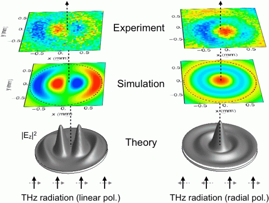
                                       Figure. When a THz wave illuminates a metallic disc surface plasmon waves
                                       are launched around its circumference propagating towards its center where
                                       they interfere at a common focus. For illumination by a linearly polarized wave
                                       a two-lobe structure is formed with a zero at the center (left panel). Using 
                                       radial polarization instead, results in optimal focusing of the surface waves 
                                       forming a strong and symmetric focal spot (right panel).

                                     
    Stefan Waselikowski1,2, Christian Fischer2, Jan Wallauer1,2 and Markus Walther1,2,3
    http://iopscience.iop.org/1367-2630/15/7/075005
    markus.walther@physik.uni-freiburg.de                            
    1 Freiburg Materials Research Center, University of Freiburg, Stefan-Meier-Strasse 21, D-79104 Freiburg, Germany
    2 Department of Molecular and Optical Physics, University of Freiburg, Stefan-Meier-Strasse 19, D-79104 Freiburg, Germany
    3 Author to whom any correspondence should be addressed

    Optimal focusing of surface plasmon polaritons in the center of a metal disc illuminated by radially polarized terahertz pulses is demonstrated. By matching the cylindrical symmetry of the metal structure with the radially polarized terahertz field, surface plasmons are excited along its entire circumference. Constructive interference in the disc center produces a sharp frequency-dependent focal spot well described by a zero-order Bessel function. We map the field distributions on the disc by terahertz (THz) near-field microscopy and compare our results with numerical simulations. For comparison, the behavior of the plasmonic lens under linearly polarized THz illumination is also characterized. The remarkable focusing capabilities of such a plasmonic lens together with its simple structure offer considerable potential for THz sensing and imaging applications.


    GENERAL SCIENTIFIC SUMMARY
    Introduction and background. Spectrocopy and imaging with terahertz (THz) radiation holds great promise for potential applications, e.g. in (bio-)molecular sensing, security screening or quality control. In order to increase spectroscopic sensitivity for THz sensing, or spatial resolution for THz imaging, it is of great importance to be able to confine the radiation to small volumes or focal spots. Unfortunately, THz field confinement is usually limited to millimetres due to the large wavelength of THz radiation (hundreds of micrometers to millimetres). Focusing THz surface-plasmon-polaritons (bounded electromagnetic waves propagating along conducting surfaces) instead of free-space radiation allows to confine fields to a small spot close to a surface, thereby potentially breaking the diffraction-limit.

    Main results. We demonstrate focusing of THz radiation in the centre of simple metal discs. By using radially polarized radiation instead of linear polarization, optimal plasmonic focusing is achieved, since the radially polarized waves perfectly match the symmetry of the circular focusing element.
    Wider implications. The remarkable focusing capabilities of such a plasmonic lens together with its simple structure offer considerable potential for achieving high sensitivities in THz sensing, as well as high spatial resolution in THz imaging applications.

    Monday, July 8, 2013

    Abstract-Optimal plasmonic focusing on a metal disc under radially polarized terahertz illumination


    Stefan Waselikowski1,2, Christian Fischer2, Jan Wallauer1,2
    and Markus Walther1,2,3
    1 Freiburg Materials Research Center, University of Freiburg,
    Stefan-Meier-Strasse 21, D-79104 Freiburg, Germany
    2 Department of Molecular and Optical Physics, University of Freiburg,
    Stefan-Meier-Strasse 19, D-79104 Freiburg, Germany
    E-mail: markus.walther@physik.uni-freiburg.de
    New Journal of Physics 15 (2013) 075005 (13pp)
    Received 7 March 2013
    Published 3 July 2013
    Online at http://www.njp.org/
    doi:10.1088/1367-2630/15/7/075005

    Optimal focusing of surface plasmon polaritons in the center of a
    metal disc illuminated by radially polarized terahertz pulses is demonstrated.
    By matching the cylindrical symmetry of the metal structure with the
    radially polarized terahertz field, surface plasmons are excited along its
    entire circumference. Constructive interference in the disc center produces a
    sharp frequency-dependent focal spot well described by a zero-order Bessel
    function. We map the field distributions on the disc by terahertz (THz) nearfield
    microscopy and compare our results with numerical simulations. For
    comparison, the behavior of the plasmonic lens under linearly polarized THz
    illumination is also characterized. The remarkable focusing capabilities of such
    a plasmonic lens together with its simple structure offer considerable potential
    for THz sensing and imaging applications

    Friday, May 24, 2013

    Abstract-Terahertz Localized Surface Plasmon Resonances in Coaxial Microcavities


    http://onlinelibrary.wiley.com/doi/10.1002/adom.201300021/abstract

    1. Withawat Withayachumnankul1,2,*, 
    2. Charan Manish Shah2, 
    3. Christophe Fumeaux1,
    4. Korbinian Kaltenecker3,4, 
    5. Markus Walther3, 
    6. Bernd M. Fischer1,4, 
    7. Derek Abbott1,
    8. Madhu Bhaskaran2, 
    9. Sharath Sriram2,*

    Coaxial microcavities etched into the surface of a doped silicon substrate are shown to support localized surface plasmon resonances at terahertz frequencies. The underlying mechanism involves coupling freely propagating terahertz waves with surface plasmon polaritons (SPPs), which propagate in a coaxial mode along the cavity walls in the axial direction. A Fabry–Pérot resonance is built up when the SPP wavenumber appropriately relates to the cavity depth. Owing to the Ohmic loss of the silicon at terahertz frequencies, the energy of the resonating SPPs is largely dissipated, leading to a modified reflection spectrum. Strong field enhancement is observed inside the cavities at resonance. The theoretical analysis is supported by numerical and experimental results. This study is a promising pathway for development of terahertz devices with applications in the areas of photonic integrated circuits, molecular sensing, and subwavelength imaging.

    Friday, December 2, 2011

    Suspended Core Subwavelength Plastic Fibers for THz Guidance


    Bora Ung, Mathieu Rozé, Anna Mazhorova, Markus Walther and Maksim Skorobogatiy

    figureExamples of a suspended solid core subwavelength fiber (a) and suspended porous core subwavelength fiber (b). (c) Output near-field distribution of the suspended fiber in (a).
    The past decade has shown significant progress in the generation and detection of terahertz (THz) signals. The first generation of fully integrated THz imaging and spectroscopy systems are being commercialized for industrial and scientific applications. These systems are often based on expensive and bulky free-space optical components that require careful alignment. In order for THz systems to become more practical, their footprint must be reduced while their ease of operation and working standoff distance must be increased. This is why researchers have emphasized the development of THz waveguides for flexible, low-loss light delivery. We report on the design and realization of a practical, single-mode, fully encapsulated low-loss THz fiber.
    It is well known that most materials in the THz exhibit high absorption losses (>0.1-20 dB/cm) that complicate the design of waveguides. Most current dielectric-based THz fibers are made of plastics since they are relatively low loss and low cost, and they offer low-temperature thermoforming and a wide array of chemical formulations.1
    The most successful designs of dielectric THz waveguides allow mode confinement to be predominantly inside the low-loss and low-dispersion gaseous regions, thus alleviating the large material losses and chromatic dispersion experienced in the THz spectrum.
    Specifically, subwavelength-size polymer fibers—featuring either a plain or porous core—have demonstrated single-mode guidance and attenuation losses in the 0.01 cm-1range.2 However, the guided mode of a subwavelength fiber features the strong presence of the fields in the cladding region, which prevents direct fiber handling due to the strong field overlap between the THz signal and the environment. This limitation represents a serious impediment to the deployment of fiber-based THz devices such as fiber endoscopes.
    To solve this problem, we fabricated a polyethylene microstructured fiber featuring a subwavelength-size core suspended by very thin bridges in the middle of an air-filled tubular jacket.3,4 Two types of fibers were successfully fabricated—one with a solid core (a) and the other with a porous core (b).
    Near-field THz microscopy measurements5 of the fiber output facet (c, top) indicate that, in the frequency range 0.28-0.48 THz, the core-guided mode of a suspended core fiber remains well confined within the tubular fiber jacket, and thus it is efficiently shielded from the environment. The experimental modal field distributions were also well reproduced by the finite-element simulations (c, bottom)—which also confirmed the effectively single-mode guidance in this frequency range.
    Moreover, the hollow region in the immediate vicinity of the core can be used as a micro-enclosure purged with dry gases, thus allowing us to forgo the cumbersome purging cages typically used in THz setups. Cutback measurements performed on the 50-cm-long fiber established low-loss guidance with signal attenuation below 0.02 cm–1 in the range of 0.28-0.48 THz.
    In summary, this work shows that all-polymer suspended core fibers enable low-loss guidance, convenient handling and mode isolation from external perturbations. Moreover, the sealed tube cladding can also be used as a purging micro-enclosure. These crucial features make polymer suspended core fibers excellent candidates for practical THz signal delivery in next-generation THz imaging and spectroscopy setups.

    Bora Ung Mathieu Rozé, Anna Mazhorova and Maksim Skorobogatiy are with the Ecole Polytechnique de Montreal, Canada. Markus Walther is with the University of Freiburg, Germany.

    References and Resources

    1. Y.S. Jin et al. J. Korean Phys. Soc. 49, 513 (2006).
    2. A. Dupuis et al. Opt. Express 18, 13813 (2010).
    3. M. Rozé et al. Opt. Express 19, 9127 (2011).
    4. B. Ung et al. CLEO:2011, paper CThN4.
    5. A. Bitzer et al. Appl. Opt. 49, E1-6 (2010)