Showing posts with label Georgia Institute of Technology. Show all posts

Thursday, February 22, 2018

In Picasso’s Blue Period, Scanners Find Secrets He Painted Over


Using tools originally developed for medicine, manufacturing and geology, researchers were able to discover new insights into “La Miséreuse accroupie,” a 1902 painting by Pablo Picasso from his “Blue Period.” CreditArt Gallery of Ontario/Picasso Estate

Scientists used a variety of tools originally developed for medicine, manufacturing
and geology to discover hidden details in the artist’s paintings and sculptures.

By 
https://www.nytimes.com/2018/02/20/science/picasso-blue-period-scans.html
Bits of color were peeking out through cracks in the dark shades of “La Miséreuse accroupie,” a 1902 painting by a young Pablo Picasso during his “Blue Period.”
That was not surprising. X-ray images taken a quarter- century ago had shown that Picasso had painted this work, known in English as “The Crouching Woman,” over another artist’s landscape.
Sandra Webster-Cook, a conservator of paintings at the Art Gallery of Ontario in Toronto, which owns the painting, also observed textures of the brush strokes that seemed neither to reflect Picasso’s composition nor the underlying landscape. “It was clear there was something else going on underneath,” Ms. Webster-Cook said.
The Ontario staff reached out to experts at the National Gallery of Art, Northwestern University and the Art Institute of Chicago.
X-ray radiography revealed a hidden landscape. Picasso rotated the canvas by 90 degrees, then painted directly over the earlier painting by another artist, not yet identified. CreditArt Gallery of Ontario

Using tools originally developed for medicine, manufacturing and geology, the researchers peered through the canvas without damaging it. They saw how Picasso had incorporated the contours of hills from the earlier painter’s landscape into the curves of the woman’s back. “Kind of a jazz riff back and forth,” said Marc Walton, a research professor of materials science and engineering at Northwestern.

The analysis also uncovered Picasso’s repeated efforts to paint the woman’s right arm. He ultimately abandoned that part of the composition, covering it with a cloak.
“So this again is getting into the mind of the artist and understanding his creative process,” Dr. Walton said.
The findings were presented Saturday at a meeting of the American Association for the Advancement of Science in Austin, Texas. At the meeting, researchers also presented new insights into Picasso’s bronze sculptures based on an analysis of the alloys.
“We’re really opening a new era of inquiry in the way these iconic works of art were made,” said Francesca Casadio, executive director of conservation and science at the Art Institute of Chicago.
Dr. Casadio and Dr. Walton are directors of the Center for Scientific Studies in the Arts, a collaboration between the university and the art institute to apply technology to art history.
Three years ago, Ms. Webster-Cook and Kenneth Brummel, the Ontario museum’s assistant curator of modern art, attended a conference in Barcelona about the scientific analysis of other Blue Period paintings. One of the scientists Ms. Webster-Cook met there was John K. Delaney, a senior imaging scientist at the National Gallery of Art in Washington.
Emeline Pouyet, left, of Northwestern University, and Sandra Webster-Cook, right, of the Art Gallery of Ontario, with “La Miséreuse accroupie” and the X-ray fluorescence instrument used to scan it. CreditArt Gallery of Ontario
Dr. Delaney later went to Ontario to examine “La Miséreuse accroupie” using a technique that records the brightness of reflected light across a swath of the visible and infrared spectrum of light.
Various molecules absorb certain colors of light. The technique, called reflectance hyperspectral imaging, allows scientists to identify minerals based on patterns of dark lines in the spectrum. It is the same technique that NASA’s Mars Reconnaissance Orbiter uses to figure out the makeup of Martian rocks from orbit.
Dr. Delaney’s images revealed the hidden right arm.
Scientists from the Center for Scientific Studies in the Arts then viewed the painting, using a portable instrument that bathed the canvas with X-rays, some of which were absorbed by elements in the painting’s pigments, then re-emitted. Different elements in the pigments radiate at different wavelengths.
Maps of the iron and chromium in the painting — Prussian blue is an iron-based pigment and chromium is used in yellow pigment — largely matched the structure of the current painting. But the patterns of cadmium, used for an array of yellows, oranges and reds, and lead, for a white pigment, showed a different painting, adding detail about the right arm and hand.
The distribution of various elements revealed in different paint layers.CreditNorthwestern University/Art Institute of Chicago

“The arm is a very strange pose,” Mr. Brummel said. “The elbow rests on the thigh, and you have this hand that’s awkwardly positioned just below the right shoulder, holding a disk.” (The disk appears to be a loaf of bread.)
Mr. Brummel said the hidden arm is similar to that of a woman in a Picasso watercolor that was also painted in 1902.
Mr. Brummel said he had made progress in identifying the original landscape artist. And he determined the scene depicted — the Labyrinth Park of Horta in Barcelona. None of the new findings would have been possible without the scans, he said.

“These scientific instruments and these collaborations with chemists and with imaging scientists are making transformational contributions to the discipline of art history,” Mr. Brummel said.
The scientists at Northwestern and the Art Institute used the same X-ray technique to examine 39 of Picasso’s bronze sculptures, cast between 1905 and 1959, and 11 painted sheet metal sculptures from the 1960s, all from the collection of the Musée Picasso in Paris.
“The tools are the same,” Dr. Casadio said. “The questions are different.”
Based on the composition of the bronze alloy, scientists were able to determine that this Picasso sculpture, “Head of a Woman, in Profile,” was cast in 1941 by the foundry of Émile Robecchi in Paris.CreditMusée national Picasso – Paris, (C) RMN-Grand Palais (Musée national Picasso-Paris), (C) Succession Picasso 2018
Photo
Over the past decade, the researchers have analyzed about 350 bronze sculptures from the late 1800s to the middle of the 20th century made by different artists in Paris. The composition of the bronze alloys provides clues about where and when the sculptures were cast.
For example, the researchers were able to figure out that the foundry of Émile Robecchi in Paris produced five of Picasso’s World War II-era bronzes that lacked an identifying stamp.
The composition of the alloys used by the Robecchi foundry in 1941 and 1942 varied significantly — some higher in tin, others higher in zinc — possibly reflecting the limited access to metals during the war.
“At the same time in Paris, the Germans were forcing private individuals and the French government to basically melt down sculptures in the city to retrieve the metal,” Dr. Casadio said.
For a later sheet metal sculpture, “Head of a Woman,” the researchers discovered that Picasso had used silver for the hair, eyes and other facial features, a perplexing choice because he then covered the expensive metal with paint.
“We took, I think, 25 additional measurements to just be a hundred percent sure,” Dr. Casadio said.
Other scientists are developing other new ways to scan artwork. In a paper published in November in the journal Scientific Reports, researchers at the Georgia Institute of Technology described how they used a form of radar to study a 17th century painting.
With a commercial terahertz scanner — industrial uses include detecting flaws in plastics — the researchers examined “Madonna in Preghiera,” by the workshop of Giovanni Battista Salvi, an Italian painter.
The painting, “Madonna in Preghiera,” seen in reflected terahertz radiation. CreditDavid S. Citrin

Terahertz waves — a form of light with wavelengths longer than infrared but shorter than microwaves — pass effortlessly through the pigments but bounce off the boundaries between different layers of paint.
Until now, terahertz waves have had limited use in the study of paintings, because the wavelengths are too long to study the thin layers. “All the echoes lie on top of each other,” said David S. Citrin, a professor of electrical and computer engineering at Georgia Tech.
Essentially, you cannot measure things smaller than the ruler you’re using.
But in this application, Dr. Citrin and his colleagues knew the echoes were bouncing off flat layers. Advanced signal processing techniques originally developed for the petroleum industry allowed them to spot layers just 20 microns thick — less than one-thousandth of an inch.
Their technique could allow other art historians to study the stratigraphy of paintings in the same way that seismologists study the structure of underground rock formations.
Dr. Walton of Northwestern said he would like to return to “La Miséreuse accroupie” with a terahertz scanner as well.
Using a wide array of scans will provide a more complete picture. “That’s really the holy grail of where we’re going with all this research,” Dr. Walton said. “By combining all these techniques together, we’re starting to get a better understanding of the entire painted structure.”

Tuesday, January 30, 2018

Optical Rectennas Get an Efficiency Boost


https://www.photonics.com/Article.aspx?AID=63054

ATLANTA, Jan. 29, 2018 — The research team that announced the first optical rectenna in 2015 reports that it has improved multiwall carbon nanotube (MWCNT) rectennas by creating and optimizing new diode structures that allow optical rectification with air-stable devices. The incorporation of double-insulator layer tunnel diodes, fabricated on MWCNT arrays, enables the use of air-stable top metals with excellent asymmetry for rectification applications. 

To provide ease of electron flow and thus a low work function, researchers from Georgia Institute of Technology (Georgia Tech) initially used calcium as the metal in the oxide insulator-metal diode junction of the rectenna. Because calcium breaks down rapidly when exposed to air, the device had to be encapsulated during operation and fabricated in a glovebox, making it difficult to fabricate and impractical for most applications. 

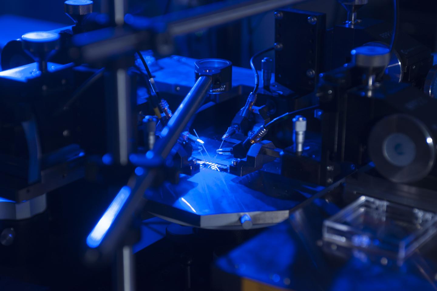
Optical rectenna, new version announced 2018, Georgia Tech.


Georgia Tech researchers have developed a new higher efficiency rectenna design. Here, the device’s ability to convert blue light to electricity is tested. Courtesy of Christopher Moore, Georgia Tech.


Researchers replaced the calcium with aluminum and experimented with a variety of oxide materials before selecting a bilayer material composed of alumina (AL2O3) and hafnium dioxide (HfO2). They created the combination coating for the carbon nanotube junction using an atomic deposition process. 

By engineering the oxide electronic properties instead of the metals, researchers were able to develop a coating that could provide the electron tunneling properties required to allow the use of air stable metals with higher work functions than calcium. Rectennas fabricated with the new combination have remained functional for as long as a year. 

The researchers also engineered the slope of the hill down which the electrons fall in the tunneling process, increasing the efficiency of the device and allowing a variety of oxide materials to be used. 

The new design increased the asymmetry of the diodes, which boosted efficiency. For the most asymmetric device structure, AL2O3-HfO2 (4/4 nm), optical rectification at a frequency of 470 THz (638 nm) was demonstrated. 

“By working with the oxide electron affinity, we were able to increase the asymmetry by more than ten-fold, making this diode design more attractive,” said professor Baratunde Cola. “That’s really where we got the efficiency gain in this new version of the device.” 

Optical rectennas operate by coupling the light’s electromagnetic field to an array of MWCNTs whose ends have been opened. The array serves as an antenna. The electromagnetic field creates an oscillation in the antenna, producing an alternating flow of electrons. When the electron flow reaches a peak at one end of the antenna, the diode closes, trapping the electrons. It then reopens to capture the next oscillation, creating a current flow. 

The switching must occur at terahertz frequencies to match the light. The junction between the antenna and diode must provide minimal resistance to electrons flowing through it while it is open, yet prevent leakage while closed. 

“The name of the game is maximizing the number of electrons that get excited in the carbon nanotube, and then having a switch that is fast enough to capture them at their peak. The faster you switch, the more electrons you can catch on one side of the oscillation,” Cola said. 

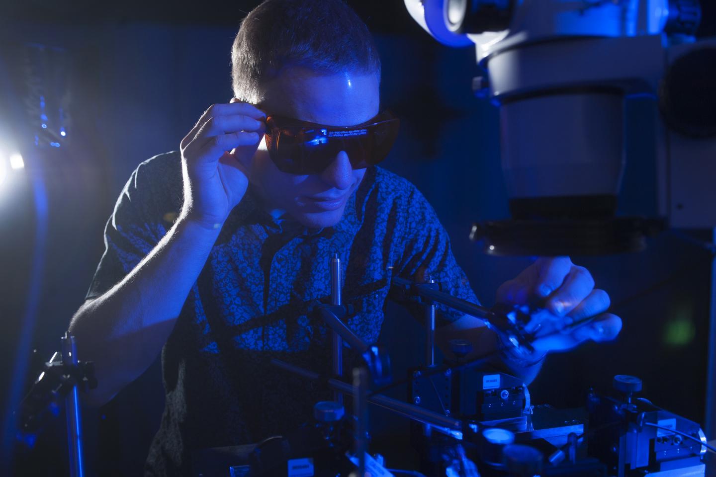
NSF Graduate Research Fellow Erik Anderson tests the conversion of blue light to electricity with a new higher efficiency rectenna design. Optical rectenna, Georgia Tech.


NSF Graduate Research Fellow Erik Anderson tests the conversion of blue light to electricity with a new higher efficiency rectenna design. Courtesy of Christopher Moore, Georgia Tech.


The early version of the rectenna produced power at microvolt levels. The new version of the rectenna produces power in the millivolt range. Conversion efficiency has gone from 10-5 to 10-3 — still low, but a significant gain. 

“Though there still is room for significant improvement, this puts the voltage in the range where you could see optical rectennas operating low-power sensors,” Cola said. “There are a lot of device geometry steps you could take to do something useful with the optical rectenna today in voltage-driven devices that don’t require significant current.” 

Cola believes the rectennas could be useful for powering internet of things devices, especially if they can be used to produce electricity from scavenged thermal energy. The researchers believe their device design could eventually compete with conventional photovoltaic technologies for producing electricity from sunlight and other sources. 

The team plans to improve its understanding of how the rectenna works, allowing further optimization. One day, Cola hopes the devices will help accelerate space travel, producing power for electric thrusters that will boost spacecraft. 

“Our end game is to see carbon nanotube optical rectennas working on Mars and in the spacecraft that takes us to Mars,” he said. 

The research was published in Advanced Electronic Materials (doi: 10.1002/aelm.201700446).

Friday, December 1, 2017

Imaging Technique Unlocks the Secrets of 17th Century Artists


Credit: Georgia Tech Photo: John Toon
Georgia Tech professor David Citrin is shown with images produced by a terahertz imaging technique. Researchers studied a 17th century painting using a terahertz reflectometry technique to analyze individual paint layers.

http://www.newswise.com/articles/imaging-technique-unlocks-the-secrets-of-17th-century-artists


Newswise — The secrets of 17th century artists can now be revealed, thanks to 21st century signal processing. Using modern high-speed scanners and the advanced signal processing techniques, researchers at the Georgia Institute of Technology are peering through layers of pigment to see how painters prepared their canvasses, applied undercoats, and built up layer upon layer of paint to produce their masterpieces.
The images they produce using the terahertz scanners and the processing technique – which was mainly developed for petroleum exploration – provide an unprecedented look at how artists did their work three centuries ago. The level of detail produced by this terahertz reflectometry technique could help art conservators spot previous restorations of paintings, highlight potential damage – and assist in authenticating the old works.
Beyond old art, the nondestructive technique also has potential applications for detecting skin cancer, ensuring proper adhesion of turbine blade coatings and measuring the thickness of automotive paints. The study was reported November 8 in the journal Scientific Reports.
“This technique allows us to see at high resolution what is beneath the surface of a painting, to assess in depth what kind of technique has been used, and to determine what defects may be present,” said Alexandre Locquet, an adjunct professor in Georgia Tech’s School of Electrical and Computer Engineering and a researcher at the Georgia Tech-CNRS international laboratory in Metz, France. “Using this, we can get information that art historians previously did not have, and we can provide information that may be helpful to the conservation and restoration of these old paintings.”
The researchers studied the painting “Madonna in Preghiera” by the workshop of Giovanni Battista Salvi da Sassoferrato, which was on loan from the Musée de la Cour d’Or, Metz Métropole, France. The examination began by placing the artwork face down on a gantry device designed to support the canvas without sagging.
Using a commercial terahertz scanner, the painting was then examined approximately every 200 microns by pulses of terahertz radiation. The scanner consists of an electromagnetic wave generator, which emits signals that penetrated through successive layers of the painting. Portions of the beam reflected back from the paint, producing signals from each layer as the scanner moved across the painting in a raster pattern similar to that used to create television images.
A computer using a signal processing technique known as sparsity-based time-domain deconvolution then processed the data, separating the signals reflected by each layer to construct a three-dimensional map of the image. The canvas support, ground, imprimatura, underpainting, pictorial and varnish layers were identified, along with a previously unknown restoration of the varnish.
“Our technique is similar to the way in which seismology can be used to identify the various layers of rock in the ground,” said David Citrin, a professor in the Georgia Tech School of Electrical and Computer Engineering. “In that case, seismologists send in an acoustic pulse and then measure the resulting echoes. In a similar way, we use a pulse of electromagnetic radiation at a frequency of around one terahertz and then look at the reflections off the various layers, a science known as stratigraphy.”
Without the signal processing, researchers might only be able to identify layers 100 to 150 microns thick. But using the advanced processing, they can distinguish layers just 20 microns thick. Paintings done before the 18th century have been challenging to study because their paint layers tend to be thin, Citrin said. Individual pigments cannot be resolved by the technique, though the researchers hope to be able to obtain that information in the future.
“This is really quite significant, because for years people have tried to use raw data, but you really can’t see much in that without processing the signals,” he said. “It takes coupling the terahertz signals with the signal processing to really make a difference.”
Terahertz radiation, also known as submillimeter radiation, operates at tremendously high frequencies. It can easily penetrate layers of paint, though it can be blocked by conductive pigments such as carbon black. The terahertz imaging technique can supplement conventional art analysis techniques such as X-rays, nuclear magnetic resonance imaging, and optical imaging.
The research team, which included graduate student Junliang Dong and collaborator Marcello Melis, has also studied other paintings, and plans to image a small part of a 12th century wood panel painting. That work will be challenging because the paint is thin and the wood surface damaged.
Citrin believes the study is the first to detect individual paint layers in a pre-18th century work of art.
“Different techniques provide different information that could be useful to art conservators and historians,” he said. “Terahertz gives us the combined ability to image a large object relatively quickly and inexpensively. We have shown that you don’t need a fancy system to extract useful information.”
Beyond paintings, Citrin’s research group has also imaged a Byzantine coin through a thick layer of oxidation, and is attempting to read an inscription in a medieval lead funerary cross also obscured by an oxide layer. They are also collaborating with a research group in Hong Kong to use the technique for characterizing the layers of skin for skin cancer detection and with another group for measuring damage in composite materials.
“Terahertz imaging is still an emerging field that has to find its best applications,” said Locquet. “We are hoping to contribute to that, and are pleased to apply science and engineering to support the humanities.”
CITATION: Junliang Dong, Alexandre Locquet, Marcello Melis & D. S. Citrin, “Global mapping of stratigraphy of an old-master painting using sparsity-based terahertz reflectometry,” (Scientific Reports, 2017) http://dx.doi.org/10.1038/s41598-017-15069-2

Saturday, April 1, 2017

What is the internet of things at nanoscale?


What is the internet of nanoscale things?

Nanoscale technology is enabling the development of devices as small as one to a few hundred nanometers (10^-9 meters). To give a sense of scale, a strand of human DNA is roughly 2.5 nanometers in diameter. At this scale, a nanomachine is defined as the most basic functional unit and able to perform simple tasks such as sensing or actuation.
Coordination and information sharing among several nanomachines will expand the potential applications of individual devices both in terms of complexity and range of operation, according to the Georgia Institute of Technology. The resulting nano-networks will be able to cover larger areas, and reach hard-to-reach locations. Moreover, the interconnection of nanoscale devices with classical networks and the internet defines a new networking paradigm, to which Georgia Institute of Technology refers to as the “internet of nano-things.”
Use cases

Some potential applications include:
In-body networks monitoring real-time blood, sickness and breath tests;
Use in public locations to monitor the spread of viruses and diseases; and
Hooked up to wearable health and environmental trackers.
When it arrives, the internet of nanoscale things could provide much more detailed, inexpensive and up-to-date pictures of our cities, homes, factories – even our bodies. Today traffic lights, wearables or surveillance cameras are getting connected to the internet with  billions of expected nanosensors harvesting huge amounts of real-time information and beaming it up to the cloud, according to Scientific American.
Methods of communication

It is still not clear how nanomachines are going to communicate. Georgia Tech presents two main alternatives for communication in the nanoscale, namely molecular communication and nano-electromagnetic communication:
Molecular communication:
This is defined as the transmission and reception of information encoded in molecules. Molecular transceivers are expected to be easily integrated in nano-devices due to their size and domain of operation. These transceivers are able to react to specific molecules, and to release others as a response to an internal command or after performing some type of processing.

Nano-electromagnetic communication:
This is defined as the transmission and reception of electromagnetic radiation from components based on novel nanomaterials.
The unique properties observed in these materials will decide the specific bandwidth for emission of electromagnetic radiation, the time lag of the emission and the magnitude of the emitted power for a given input energy.
Network architecture for IoNT

Georgia Tech proposes the study of the terahertz band for nano-electromagnetic communication and provides a network architecture for nano devices.
In intrabody networks, nanomachines such as nanosensors and nanoactuators deployed inside the human body are remotely controlled from the macroscale and over the internet by an external user such as a health care provider. The nanoscale is the natural domain of molecules, proteins, DNA, organelles and the major components of cells. Amongst others, existing biological nanosensors and nanoactuators provide an interface between biological phenomena and electronic nano-devices, which can be exploited through this new networking paradigm.
In the interconnected office, every single element normally found in an office and even its internal components are provided of a nanotransceiver which allows them to be permanently connected to the internet.
The use cases in these different environments shows that nanotechnology has the ability to create new applications in the biomedical, industrial and military fields as well as in consumer and industrial goods.
Demands for nano IoT

These are factors, according to Georgia Tech, that will increase demand for nano devices:
Convenience and almost seamless deployment;
Tiny and nonobtrusive devices;
The possibility to harvest vibrational, mechanical or even electromagnetic energy from the environment;
Ultra-low power consumption; and
Reasonable computing capabilities.
Here are the physical components required for the internet of nano things architecture:
Nano-nodes
The smallest and simplest nanomachines, they are able to perform simple computation, have limited memory and can only transmit over very short distances, mainly because of their reduced energy and limited communication capabilities. Biological nanosensor nodes inside the human body and nanomachines with communication capabilities integrated in all types of things such as books, keys, or paper folders are good examples of nano-nodes.
Nano-routers

Comparatively larger computational resources than nano-nodes and are suitable for aggregating information coming from limited nanomachines. In addition, nano-routers also can control the behavior of nano-nodes by exchanging very simple control commands (on/off, sleep, read value, etc.). However, this increase in capabilities involves an increase in their size, and this makes their deployment more invasive. Nano-micro interface devices are able to aggregate the information coming from nanorouters, to convey it to the microscale, and vice versa.
Gateway

Enables the remote control of the entire system over the internet. For example, in an intrabody network scenario, an advanced cellphone can forward the information it receives from a nano-micro interface in our wrist to our health care provider. In the interconnected office, a modem-router can provided this functionality. Despite the interconnection of microscale devices, the development of gateways and the network management over the internet are still open research areas, in the remaining of this article we mainly focus on the communication challenges among nanomachines.
Technologies enabling smaller data collection

Scientists have started shrinking sensors from millimeters or microns in size to the nanometer scale, small enough to circulate within living bodies and to mix directly into construction materials. There are five new developments that are helping enable the shrinking of sensors and collection of data from nano devices, according to Computer Business Review.
Nanotubes

Carbon nanotubes are a nanotechnology constructed with a length-to-diameter ratio of up to 132,000,000:1.
Uses of the solution span from incorporation in portable electronics to help fighting cancer and creating artificial muscles.
Bleeding plastic

Scientists have also developed a bleeding plastic with self-healing capabilities that could put an end to nearly anything getting broken, including cars, airplanes or everyday devices.
Nano-nodes

Nano-nodes are nanomachines with the capability to perform simple computation, but could be used in the future to make nearly every object and person connected to the internet.
In a whitepaper from IEEE Wireless Communications, Ian Akyildiz and Josep Jornet from the Georgia Institute of Technology explained that nano-nodes have limited memory, and can only transmit over very short distances, mainly because of their reduced energy and limited communication capabilities.
Nanoantennas

Nanoantennas are a new emerging technology that could help power wearables, smarten up buildings or keep lights on.


Tuesday, February 28, 2017

OT-Mass Spectrometry Gets a New Power Source and a New Life


Image: Georgia Institute of Technology/Nature Nanotechnology


Mass spectrometry is a chemical analysis and detection tool that has been around for 130 years. In that time there have been so many tweaks and improvements that observers have become a bit blasé about the next big leap in its development.
But the latest improvement out of the Georgia Institute of Technology may be the biggest yet for the venerable old analytical tool. In research described Nature Nanotechnology, the Georgia Tech researchers have managed to make mass spectrometry more sensitive than ever before, more portable, cheaper and even safer. All of these advancements were accomplished by replacing the direct current power source typically used as power source with triboelectric nanogenerators (TENGs). You can see a demonstration of the technology at work in the video below.
While the researchers concede the mechanism by which the enhancement takes place demands more investigation, they believe the unique aspects of the TENG output—oscillating high voltage and controlled current—should enable improvements in the ionization process, increasing the voltage applied without damaging samples.
The so-called TENGs were developed at Georgia Tech back in 2012 and Zhong Lin Wang and his colleagues at Georgia Tech have been improving the technology and expanding its applications ever since. TENGs essentially harvest static electricity from friction. The TENG devices consist of two different materials that are rubbed together. In this way, materials that tend to give off electrons, such as glass or nylon, will donate them to materials that tend to absorb them, such as silicon or teflon. By converting mechanical energy from friction to electricity the TENGs can power small electronic devices.
Applied to mass spectrometry, the TENGs replace the direct current power source for generating the ions. This takes the advantage of a fixed input charge in each cycle of the operation of the TENG regardless of the current or voltage, allowing the mass spectrometer to analyze the smallest possible sample at the highest possible sensitivity.
“The sensitivity has been increased to being able to detect down to 100 molecules,” said Wang in an e-mail interview with IEEE Spectrum. “This is the highest ever.”
Wang also points out how efficient the TENG power source is in using samples. In mass spectrometry, a sample is ionized and the ions are sorted according to their mass-to-charge ratio. But with DC voltages, the number of generated ions does not depend on the applied voltage in a straightforward fashion. As a result, controlling the number of charges used in the ionization of neutral species is usually impossible. The fixed number of charges provided by TENGs offer unprecedented control over ion generation. This makes it very efficient in how it uses the sample.
“The key here is that the total charge delivered in each cycle is entirely controlled and constant regardless of the speed at which the TENG is triggered,” said Wang in a press release.
Facundo Fernández, a professor in Georgia Tech’s School of Chemistry and Biochemistry, added: “Our discovery is basically a new and very controlled way of putting charge onto molecules. We know exactly how much charge we produce using these nanogenerators, allowing us to reach sensitivity levels that are unheard of – at the zeptomole scale (10-21th part of a mole or about 600 molecules). We can measure down to literally hundreds of molecules without tagging.”
The Georgia Tech team has measured the TENGs generating as much as 6000 and 8000 volts as a mass spectrometry ionizer. Standard ionizers normally operate at less than 1500 volts.
“Because the voltage from these nanogenerators is high, we believe that the size of the sample droplets can be much smaller than with the conventional way of making ions,” Fernández said. “That increases the ion generation efficiency. We are operating in a completely different electrospray regime, and it could completely change the way this technology is used.”
By eliminating the often high-voltage power supplies, mass spectrometry could become more portable, leading the researchers to speculate that they could be used in extreme and harsh environments since they would become a durable self-contained unit.
While dramatically improving sensitivity, portability and adding a bit of safety by replacing the high-voltage power supplies, the TENGs also enable the deposition of ions onto surfaces, including non-conducting ones. This becomes possible because the TENGs are creating an oscillating ionization that produces a sequence of alternating positive and negative charges, resulting in a net neutral surface.
Wang added: “This opens a new field for applying TENG in portable and mobile instruments with high performance. The next phase of research is to optimize the performance of the system so that it can be used for sophisticated analytical chemistry and biochemistry.”