Showing posts with label nanotubes. Show all posts

Monday, April 20, 2020

Spectroscopic terahertz imaging probes the inner structures of 0D-3D nanomaterials

Terahertz time-domain scanning technology provides camera-less, lattice-resolution, layer-by-layer imaging and identification of the internal structures of simple and complex nanomaterials.
FIGURE 1. For the terahertz nanoscanner setup, both reflection- and transmission-mode measurements are possible (a). The sample remains stationary while the nanoscanner scans the sample over a chosen area or volume. Here, an optical wafer is mounted on the terahertz nanoscanner to prepare for imaging (b).

ANIS RAHMAN

https://www.laserfocusworld.com/detectors-imaging/article/14167860/spectroscopic-terahertz-imaging-probes-the-inner-structures-of-0d3d-nanomaterials

As nanotechnology progresses, the photonics community is now tasked with measuring and identifying nanoscale materials with extremely small and varied structural parameters.
On the nanoscale, zero-dimensional (0D) structures are those that have truly nanoscale dimensions on the order of 10 nm in size or less, such as quantum dots.1 These 0D materials do not form a molecular network of multidimensionality, but remain as a nano-dimensional entity, with a size smaller than the de Broglie wavelength.
Nanomaterials such as nanowires or nanorods that form a molecular network and extend their length along one direction only while their diameter remains unchanged at <100 nm are termed one-dimensional (1D) nanomaterials. Alternatively, two-dimensional (2D) or planar nanomaterials like graphene and carbon nanotubes form a molecular network in both the x- and y-directions, with examples of 2D allotropes including borophene, germanene, silicene, phosphorene, and stanine. And finally, when aggregated in a size bigger than 100 nm, all materials fall in the realm of 3D, expanding their unit cells in all three orthogonal directions.
While modern nanophotonic processes have facilitated the synthesis and production of these exotic nanomaterials, measurement and proper characterization of these nanoscale and subnanoscale materials pose new challenges to available instrumentation.
The TNS3DI terahertz time-domain imager from ARP (Harrisburg, PA) can analyze and quantify the above-mentioned materials, from 0D to 3D. In earlier research, the measurement of surface properties such as surface topography, texture, step height, and more—over a wide range of surfaces and materials—has been demonstrated.2 But the metrology of 0D–3D materials involves not only quantifying their size parameter and size distribution, it also involves quantifying the interaction of one nanomaterial with another.

Zero-dimensional quantum dots

A quantum dots (QD) is a nanocrystal that confines electrons and holes within its de Broglie wavelength; as such, these nanocrystals are only a few nanometers in size. At this scale, the electron is confined in a small region of space and occupies discrete energy levels that are analogous to those of a single atom. For this reason, QDs are also referred to as “artificial atoms.”
Quantum dots are most commonly fabricated using epitaxial techniques such as molecular beam epitaxy (MBE) or by colloidal chemistry. Ordinarily, an atomic force microscope (AFM) is used to image the QDs; a scanning electron microscope (SEM) can also be used when the QDs are deposited on a substrate such as a glass slide.
Alternatively, the ARP TNS3DI camera-less imaging technique characterizes 0D to 3D nanomaterials using terahertz radiation. The terahertz setup consists of a nanoscanner that digitizes an object over a 3D space (see Fig. 1).
It is well known that the resolution of images formed by a focusing lens on a recording medium—photographic film or a charge-coupled device (CCD)—is determined by the Abbe diffraction limit (ADL) that sets the resolution to the highest value of half the wavelength of the light used for imaging. Fortunately, ADL can be overcome (and higher-resolution images obtained) through terahertz multispectral reconstructive imaging.3
Since most materials are transparent to terahertz radiation, the ARP nanoscanner can probe and visualize subsurface features in a nondestructive, noncontact fashion. Together, hardware and software enable user-defined pixel sizes (voxel sizes in 3D), while a digital camera displays and records the processed signal from a sample with a fixed pixel size.
In a camera, an object is focused on a CCD or a focal plane array and a built-in processor displays the image on a screen and saves the image data in a file. In contrast, the terahertz technique eliminates the CCD and lens system by using a nanoscanner and a suitable computer algorithm for image generation and processing, eliminating the focusing lens and the CCD.
Here, the object to be imaged is scanned (digitized) along the three orthogonal axes for 3D imaging or digitized on a plane for surface imaging. A matrix containing the digitized reflected signal (or, equivalently, the transmitted signal) is recorded in a file and then processed by a suitable algorithm. Experimental measurement of the lattice constant of metallic nickel at 0.353 nm is the same value accepted in the scientific literature, therefore validating the terahertz imaging technique.
FIGURE 2. The terahertz nanoscanner reveals a low-resolution image of quantum dots (QDs) spun on a silicon wafer (a); software translates the QDs into a 3D image or data cube (b)

In the case of multiple 0D QDs spun on a silicon wafer, the nanoscanner imaging data shows the physical attributes of the QDs in the xy plane and is then translated into a 3D data cube that clearly delineates the individual dots (see Fig. 2). From the intensity matrix, a detailed image of the individual dots can be generated to obtain dimensional information (see Fig. 3).
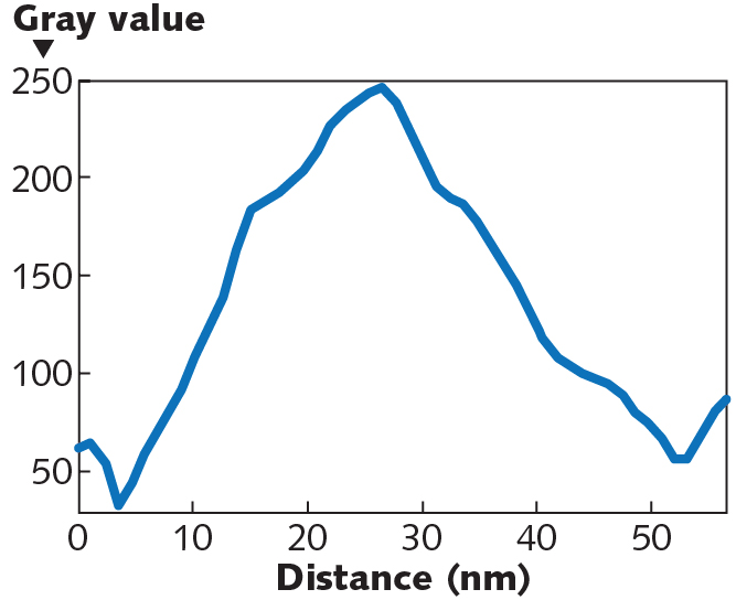
FIGURE 3. Graphical analysis of a single QD in a field of QDs (a) shows a diameter of approximately 8 nm (b).

One-dimensional structure characterization

Nanowires and carbon nanotubes are examples of 1D nanomaterials. Since their length is much bigger compared to their width (diameter), they qualify as 1D structures. Carbon nanotubes (CNTs) are cylindrical nanostructures that can be classified as single-walled and multi-walled carbon nanotubes (SWCNTs and MWCNTs).
With unique properties that lend themselves to extraordinary applications in electronics and optics, terahertz analysis with the TNS3DI can measure numerous properties. With a built-in terahertz time-domain spectrometer, it can perform both spectral analysis and deep-level spectroscopy, whereby spectra are collected at different depths of a sample as specified by the user.
The TNS3DI implements a pump-probe technique with ARP’s proprietary terahertz source that works with a patented dendrimer dipole excitation (DDE) mechanism.4 Collecting the terahertz time-domain signal over a few picoseconds generates a signal known as an interferogram and the Fourier transform of that interferogram yields the absorption spectra of the sample (see Fig. 4). Prominent absorption peaks in the spectrum are observed at 1.72, 4.29, 6.61, 13.70, and 15.59 THz for the CNT samples. Here, data was collected between 0.1 and 30 THz—however, to increase legibility, the spectrum is only displayed up to 20 THz.
FIGURE 4. Fourier transforms of the time-domain data reveal the broadband terahertz absorbance spectra of MWCNTs.

Although CCD imaging devices such as digital microscopes and cameras normally have good resolution, higher levels of resolution are challenging and only surface details can be imaged. And while transmission electron microscopy (TEM) offers high resolution, it is strictly a destructive technique with laborious sample preparation, is only for small geometries, and does not offer 3D capability. Focused ion beam and x-ray diffractive (XRD) imaging are also destructive techniques.
FIGURE 5. A 3D image of a section of a MWCNT reveals the 3D organization of the annealed CNTs (a); a single CNT’s width is shown by the circle (b).
FIGURE 6. Size analysis from the MWCNT image reveals an average individual CNT diameter of 48.54 nm (edge to edge).

In contrast, TNS3DI-based terahertz imaging defines the pixel size (or, the voxel size in 3D) by a hardware and software combination, making it possible to characterize complex structures such as MWCNTs. For example, a representative 3D image of a section of MWCNTs shows various strands of the inner material from a 2 μm × 1 μm section, revealing a single CNT’s width (see Fig. 5). Size analysis shows the average diameter of the CNT to be about 48 nm (see Fig. 6). Further analysis shows the length at about 1200 nm or 1.2 µm—a value that compares favorably with TEM, electron-diffraction, and Raman studies.5

Two-dimensional graphene

In theory, graphene is supposed to be a single layer of carbon atoms—a 2D nanomaterial. Graphene is mostly extracted from graphite—an allotrope of the element carbon meaning it possesses the same atoms, but they are arranged differently, giving the material different properties. In reality, however, graphene is an exfoliate possessing several layers of carbon sheets.
At ARP, terahertz multispectral computed imaging provides direct measurement of the graphene layers (that is, the number of sheets of graphene) in an exfoliate. It also measures the thickness of a single layer in the exfoliate. High-quality graphene is expected to have less than 10 layers in an exfoliate and should have a layer (sheet) thickness of <1 nm. Graphene oxide’s (GO) number of sheets in an exfoliate is higher and so is the thickness of each sheet.
Once the 3D image of a given exfoliate is generated, several useful parameters may be extracted. In addition to the number of layers and their thicknesses, a fast-Fourier transform (FFT) diffraction pattern can also be generated to describe crystallographic information. This volumetric imaging data not only allows layer-by-layer thickness measurement, but can also be used to obtain an FFT diffraction pattern.

Three-dimensional nanomaterials

A recently published paper by Rahman et al. in Novel Research in Sciences (NRS) investigated the dilation of nickel lattice from samples of alumina rods containing layers of metals and insulators that were subjected to what is known as a low-energy nuclear reaction (LENR) experiment.6 The samples were obtained from Brillouin Energy Corporation (Berkeley, CA), which demonstrated that the samples produced excess output energy compared to the input, under their experimental conditions.
However, Rahman et al. found a different explanation of this higher output energy than what was thought to be LENR process. The authors postulated that the higher-energy-generation effect observed in the experiment was most likely a “lattice-driven phenomenon” as opposed to the nuclear transmutation of LENR.
Rahman et al. conducted an in situ and ex situ systematic investigation that assumes a “time-crystal-like non-equilibrium” process is driving the energy balance. Time crystals—a newer concept—are states of matter whose patterns repeat at set intervals of time and space. They are systems in which time symmetry is spontaneously broken. A time crystal never reaches thermal equilibrium, as it is a type of nonequilibrium matter proposed in 2012.
FIGURE 7. In high-resolution 3D images of four samples (1 µm3), the nickel-rich area of the materials shows embedded nanograins of alumina that cause lattice deformations

Using the terahertz time-domain technique, the authors analyzed a heated sample of crystalline nickel, which is more fluid-like than rigid near the glass-transition point (see Fig. 7). The applied thermal energy in a radio-frequency field sets the fluid-like nickel lattice into oscillation, producing a nonradiative transition that creates the increased heat energy. Instead of using an electron microscope, the terahertz technique breaks ground by using a larger terahertz wave that breaks the Abbe diffraction limit for lattice imaging. The experiment proves that the LENR is actually nonexistent and rather, a time-crystal-like non-equilibrium effect is driving the energy balance for what was considered to be a LENR phenomenon.
The ARP terahertz nanoscanner brings a new dawn of CCD-less imaging as nanomaterial characterization techniques have progressed from photographic film to CCD and finally to nanoscanner-based technologies. It is hoped this new technique will aid in solving a number of problems in industry and academia alike.
REFERENCES
1. A. Rahman et al., J. Biosens. Bioelectron., 7, 3, 1–8 (2016); doi:10.4172/2155-6210.1000221.
2. A. Rahman, “Application of TNS3DI as a Surface Metrology Tool,” doi:10.13140/rg.2.2.30786.73921 (2020).
3. A. Rahman and A. K. Rahman, IEEE Trans. Semicond. Manuf., 32, 1, 7–13 (Feb. 2019); doi:10.110 9/TSM.2018.2865167.
4. A. Rahman, A. K. Rahman, and D. A. Tomalia, Nanoscale Horiz., 2, 127–134 (Mar. 20, 2017); doi:10.1039/c7nh00010c.
5. W. Ghann et al., J. Nanomed. Nanotechnol., 10, 4, 535 (2019); doi:10.35248/2157-7439.19.10.535.
6. A. Rahman et al., Nov. Res. Sci. 2, 4, NRS.000545.2019 (2019); doi:10.31031/nrs.2019.2.000545.
Anis Rahman is president and chief technology officer at Applied Research & Photonics (ARP), Harrisburg, PA; e-mail: a.rahman@arphotonic.nethttp://arphotonics.net.

Wednesday, November 29, 2017

Abstract-Polarization independent triple-band (5,4) semiconducting carbon nanotube metamaterial absorber design for visible and ultraviolet regions


Madina Obaidullah,  Volkan Esat, Cumali Sabah,

https://www.spiedigitallibrary.org/journals/Journal-of-Nanophotonics/volume-11/issue-4/046011/Polarization-independent-triple-band-54-semiconducting-carbon-nanotube-metamaterial-absorber/10.1117/1.JNP.11.046011.short?SSO=1

Various metamaterial absorber designs operating in the microwave, infrared, visible, and ultraviolet frequency regions have been proposed in the literature. However, only a few studies have been done on the metamaterials that absorb in both visible and ultraviolet solar spectra. A triple-band polarization-insensitive metamaterial absorber structure with semiconducting single-walled carbon nanotube as the dielectric layer is proposed to efficiently absorb the incident electromagnetic radiations in visible and ultraviolet frequency regions. A unit cell of this design comprises three basic components in the form of metal–semiconductor–metal layers. The metallic part of the structure is aluminum, and the (5,4) single-walled carbon nanotube is used as the semiconducting material. The electromagnetic response of the proposed design is numerically simulated in the visible and ultraviolet regions with the maximum absorption rates of 99.75% at 479.4 THz, 99.94% at 766.9 THz, and 97.33% at 938.8 THz with corresponding skin depths of 13.0, 12.8, and 12.9 nm, respectively. Thus, solar cells based on this metamaterial absorber can offer nearly perfect absorption in the suggested frequency regions. The simple configuration of the design provides flexibility to control geometric parameters to be used in the solar cell and possesses the capability to be rescaled for other solar spectrum.
© 2017 Society of Photo-Optical Instrumentation Engineers (SPIE

Tuesday, July 19, 2016

Abstract-Density Detection of Aligned Nanowire Arrays Using Terahertz Time-Domain Spectroscopy


Wenfeng Xiang, Xin Wang, Yuan Liu, JiaQi Zhang, Kun Zhao,

http://link.springer.com/article/10.1186/s11671-016-1551-1

A rapid technique is necessary to quantitatively detect the density of nanowire (NW) and nanotube arrays in one-dimensional devices which have been identified as useful building blocks for nanoelectronics, optoelectronics, biomedical devices, etc. Terahertz (THz) time-domain spectroscopy was employed in this research to detect the density of aligned Ni NW arrays. The transmitted amplitude of THz peaks and optical thickness of NW arrays was found to be the effective parameters to analyze the density change of NW arrays. Owing to the low multiple scattering and high order of Ni NW arrays, a linear relationship was observed for the transmitted amplitude and optical thickness regarding NW density, respectively. Therefore, THz technique may be used as a promising tool to characterize the density of one-dimensional structures in the large-scale integrated nanodevice fabrication.

Saturday, January 9, 2016

Abstract-Near-Field Infrared Pump–Probe Imaging of Surface Phonon Coupling in Boron Nitride Nanotubes


 Department of Chemistry, University of Toronto, Toronto, Ontario M5S 3H6, Canada
 Department of Chemistry, Lehigh University, 6 East Packer Avenue, Bethlehem, Pennsylvania 18015, United States
§ National Institute for Materials Science (NIMS), Tsukuba, Ibaraki 305-0044, Japan
J. Phys. Chem. Lett.20167, pp 289–294
DOI: 10.1021/acs.jpclett.5b02438
Publication Date (Web): January 4, 2016
Copyright © 2016 American Chemical Society


Surface phonon modes are lattice vibrational modes of a solid surface. Two common surface modes, called longitudinal and transverse optical modes, exhibit lattice vibration along or perpendicular to the direction of the wave. We report a two-color, infrared pump-infrared probe technique based on scattering type near-field optical microscopy (s-SNOM) to spatially resolve coupling between surface phonon modes. Spatially varying couplings between the longitudinal optical and surface phonon polariton modes of boron nitride nanotubes are observed, and a simple model is proposed.

Wednesday, February 5, 2014

Abstract-Mechanotunable monatomic metal structures at graphene edges


Monatomic metal (e.g. silver) structures could form preferably at graphene edges. Their structural and electronic properties are explored here by performing density functional theory based first-principles calculations. The results show that cohesion between metal atoms and electronic coupling between metal atoms and graphene edges offer remarkable structural stability. Outstanding mechanical properties of graphene allow tunable properties of the metal monatomic structures by applying mechanical loads. Moreover, metal rings and helices can form at open ends of carbon nanotubes and edges of twisted graphene ribbons. These findings suggest the role of graphene edges as an efficient one-dimensional template for low-dimensional metal structures that are mechanotunable.

Monday, December 9, 2013

Plasmons Ripple at Terahertz Frequencies in Metallic and Doped Carbon Nanotubes


Published on December 9, 2013 at 5:19 AM

Carbon nanotubes carry plasmonic signals in the terahertz range of the electromagnetic spectrum, but only if they’re metallic by nature or doped.


The ability to sort carbon nanotubes by type through a process called “density gradient ultracentrifugation (DGU)” allowed Rice researchers to test purified batches of nanotubes to find the cause of terahertz peaks in spectroscopic experiments. They determined that free electrons formed plasmons that ripple at terahertz frequencies in metallic and doped nanotubes. Courtesy of the Kono Laboratory

In new research, the Rice University laboratory of physicist Junichiro Kono disproved previous theories that dominant terahertz response comes from narrow-gap semiconducting nanotubes.
Knowing that metallic or doped nanotubes respond with plasmonic waves at terahertz frequencies opens up the possibility that the tubes can be used in a wide array of optoelectronic amplifiers, detectors, polarizers and antennas.
The work by Kono and his Rice colleagues appeared online recently in the American Chemical Society journal Nano Letters.
Scientists have long been aware of a terahertz peak in nanotubes, the tiny cylinders of rolled-up carbon that show so much promise for advanced materials. But experiments on batches of nanotubes, which generally grow in a willy-nilly array of types, failed to reveal why it was there.
The origin of the peak was not explainable because researchers were only able to experiment on mixed batches of nanotube types, said Qi Zhang, a graduate student in Kono’s group and lead author of the paper. “All the previous work was done with a mixture of semiconducting and metallic tubes. We are the first to clearly identify the plasmonic nature of this terahertz response,” he said.
Rice’s growing expertise in separating nanotubes by type allowed Kono and his group to test for terahertz peaks in batches of pure metallic nanotubes known as “armchairs” as well as nonmetallic, semiconducting tubes.
“Metallic carbon nanotubes are expected to show plasmon resonance in the terahertz and infrared range, but no group has clearly demonstrated the existence of plasmons in carbon nanotubes,” Zhang said. “Previously, people proposed one possible explanation — that the terahertz peak is due to interband absorption in the small band gaps in semiconducting nanotubes. We rejected that in this paper.”
Plasmons are free electrons on the surface of metals like gold, silver or even aluminum nanoparticles that, when triggered by a laser or other outside energy, ripple like waves in a pond. Strong waves can trigger plasmon responses in adjacent nanoparticles. They are being investigated at Rice and elsewhere for use in sophisticated electronic and medical applications.
The Kono group’s research showed plasmons rippling at terahertz frequencies only along the length of a nanotube, but not across its width. “The only way charge carriers can move around is in the long direction,” Kono said. The researchers previously used this fact to demonstrate that aligned carbon nanotubes act as an excellent terahertz polarizer with performance better than commercial polarizers based on metallic grids.
Nanotubes can be thousands of times longer than they are wide, and the ability to grow them (or cut them) to specific lengths or to dope semiconducting nanotubes to add free carriers would make the tubes highly tunable for terahertz frequencies, Kono said.
“This paper only clarifies the origin of this effect,” he said. “Now that we understand it, there’s so much to do. We will be making various terahertz devices, architectures and systems based on carbon nanotube plasmons.”
Rice alumni Erik Hároz, now a postdoctoral researcher at Los Alamos National Laboratory, and Lei Ren, a researcher at TGS, co-authored the paper with undergraduate student Zehua Jin, postdoctoral researcher Xuan Wang, senior research scientist Rolf Arvidson and Andreas Lüttge, a research professor of Earth science and chemistry, all of Rice. Kono is a professor of electrical and computer engineering and of physics and astronomy and of materials science and nanoengineering.
The Department of Energy, the National Science Foundation and the Robert A. Welch Foundation supported the research.
Source: http://www.rice.edu/

Tuesday, June 11, 2013

Abstract-Anisotropic Dielectric Relaxation of the Water Confined in Nanotubes for Terahertz Spectroscopy Studied by Molecular Dynamics Simulations


J. Phys. Chem. B, Just Accepted Manuscript
DOI: 10.1021/jp3120435
Publication Date (Web): June 10, 2013
Copyright © 2013 American Chemical Society

http://pubs.acs.org/doi/pdfplus/10.1021/jp3120435

The dynamics and structure of hydrogen-bond network in confined water are of importance in understanding biological and chemical processes. Recently, the terahertz (THz) time domain spectroscopy is widely applied for studying the kinetics of molecules and hydrogen-bond network in water. However, the characteristics of the THz spectroscopy varying with respect to the confinement and the mechanism underlying the variation are still unclear. Here, based on molecular dynamics simulations, the relationship between the anisotropic dielectric relaxation and the structure of the water confined in a carbon nanotube (CNT) was investigated. The results show that there are two preferred hydrogen-bond orientations of the confined water in the nanotube: 1) parallel to the CNT axis and 2) perpendicular to the CNT axis, which are clearly different. Moreover, the response of the orientations to the increment of the CNT diameters is opposite, leading to the opposite variations of the dielectric relaxation times along the two directions. The anisotropy in the relaxation time can be presented by the anisotropic dielectric permittivity which is able to be observed through THz spectroscopy. The anormal behaviors above are attributed to the special structure of the water close to the nanotube wall due to the confinement and hydrophobicity of CNT. These studies contribute an important step in understanding the THz experiments of water in nano scales, and designing a chamber for specific chemical and biological reactions by controlling the diameters and materials of the nanotube.

Wednesday, February 27, 2013

Nanotube photodetector built by Rice University & Sandia Labs


This illustration shows an array of parallel carbon nanotubes 300 micrometers long that are attached to electrodes and display unique qualities as a photodetector, according to researchers at Rice University and Sandia National Laboratories. Credit: Sandia National Laboratories

Read more at: http://phys.org/news/2013-02-nanotube-photodetector-built.html#jCp
http://phys.org/news/2013-02-nanotube-photodetector-built.html#jCp
Researchers at Rice University and Sandia National Laboratories have made a nanotube-based photodetector that gathers light in and beyond visible wavelengths. It promises to make possible a unique set of optoelectronic devices, solar cells and perhaps even specialized cameras.

A traditional camera is a light detector that captures a record, in chemicals, of what it sees. Modern digital cameras replaced film with semiconductor-based detectors. But the Rice detector, the focus of a paper that appeared today in the online Nature journal Scientific Reports, is based on extra-long carbon nanotubes. At 300 micrometers, the nanotubes are still only about 100th of an inch long, but each tube is thousands of times longer than it is wide. That boots the broadband detector into what Rice physicist Junichiro Kono considers a macroscopic device, easily attached to electrodes for testing. The nanotubes are grown as a very thin "carpet" by the lab of Rice chemist Robert Hauge and pressed horizontally to turn them into a thin sheet of hundreds of thousands of well-aligned tubes. They're all the same length, Kono said, but the nanotubes have different widths and are a mix of conductors and semiconductors, each of which is sensitive to different wavelengths of light. "Earlier devices were either a single nanotube, which are sensitive to only limited wavelengths," he said. "Or they were random networks of nanotubes that worked, but it was very difficult to understand why." "Our device combines the two techniques," said Sébastien Nanot, a former postdoctoral researcher in Kono's group and first author of the paper. "It's simple in the sense that each nanotube is connected to both electrodes, like in the single-nanotube experiments. But we have many nanotubes, which gives us the quality of a macroscopic device." With so many nanotubes of so many types, the array can detect light from the infrared (IR) to the ultraviolet, and all the visible wavelengths in between. That it can absorb light across the spectrum should make the detector of great interest for solar energy, and its IR capabilities may make it suitable for military imaging applications, Kono said. "In the visible range, there are many good detectors already," he said. "But in the IR, only low-temperature detectors exist and they are not convenient for military purposes. Our detector works at room temperature and doesn't need to operate in a special vacuum." The detector is also sensitive to polarized light and absorbs light that hits it parallel to the nanotubes, but not if the device is turned 90 degrees. The work is the first successful outcome of a collaboration between Rice and Sandia under Sandia's National Institute for Nano Engineering program funded by the Department of Energy. François Léonard's group at Sandia developed a novel theoretical model that correctly and quantitatively explained all characteristics of the nanotube photodetector. "Understanding the fundamental principles that govern these photodetectors is important to optimize their design and performance," Léonard said. Kono expects many more papers to spring from the collaboration. The initial device, according to Léonard, merely demonstrates the potential for nanotube photodetectors. They plan to build new configurations that extend their range to the terahertz and to test their abilities as imaging devices. "There is potential here to make real and useful devices from this fundamental research," Kono said. More information: www.nature.com/srep/2013/130226/srep01335/full/srep01335.html


Tuesday, May 22, 2012

Graphene and carbon nanotubes could lead to terahertz fast computers, phones


Graphene and carbon nanotubes could improve the electronics used in computers and mobile phones, says a researcher from the University of Gothenburg, Sweden.
Carbon nanotubes and graphene are both made up of carbon and have unique properties. Graphene comprises an atom-thick layer of carbon atoms, while carbon nanotubes can be likened to a graphene sheet that has been rolled up to form a tube.
"If you stretch a graphene sheet from end to end the thin layer can oscillate at a basic frequency of getting on for a billion times a second. This is the same frequency range used by radios, mobile phones and computers," said researcher Anders Nordenfelt.
It is hoped that the limited size and weight of these new carbon materials could further reduce both the size and power consumption of our electronic circuits.
In addition to new applications in electronics, research is under way into how graphene can be used to weigh extremely small objects such as DNA molecules.
The high mechanical resonance frequencies mean that carbon nanotubes and graphene can pick up radio signals.
"The question is whether they can also be used to produce this type of signal in a controlled and effective way. This assumes that they themselves are not driven by an oscillating signal that, in turn, needs to be produced by something else," stated Anders Nordenfelt.
In his research Nordenfelt carried out a mathematical analysis to demonstrate that it is possible to connect the nanowire with a fairly simple electronic circuit, and at the same time to apply a magnetic field and thus get the nanowire to self-oscillate mechanically.
"At the same time we're converting a direct current to an alternating current with the same frequency as the mechanical oscillation," explained Nordenfelt.
In addition to their own keynote, all mechanical strings have harmonics that, for example, give different musical instruments their own particular sound.
"An unexpected and very interesting result is that the method I've proposed can be used to get the nanowire to self-oscillate in one of its harmonics. You can change the harmonic by altering the size of one or more of the electronic components," Nordenfelt said.
In principle, there are an infinite number of harmonics with unlimited high frequencies, but there are practical limitations.
A long-held research dream is to produce signals in the terahertz range, with trillions of oscillations per second.
This area is particularly interesting as it lies on the boundary between microwaves and infrared radiation that, to date, has been the subject of relatively little research. It is an area that has been too fast for electronic circuits, but too slow for optical circuits.
"We can't get these really high frequencies with my method as things stand, but it could be something for the future," added Anders Nordenfelt. (ANI)

Monday, April 9, 2012

Scale made with a carbon nanotube sensitive enough to register a proton's mass

Schematic (upper left) and electron micrograph of a carbon nanotube resonator. The tube (the thin gray horizontal line) changes its vibrational frequency in response to added atoms and molecules.


My Note: No exciting developments I know about dealing specifically with THz, or related companies so here's a semi-related story about graphene, which holds much promise for THz in the future. 

By
On macroscopic scales, we can use the force of gravity to help us determine mass. But on microscopic scales, other forces typically dominate. Nanoscopic mechanical resonators, which change their vibrational frequency in response to added mass, have allowed us to measure smaller objects, finding the masses of biological cells and large molecules. Now, researchers at the Universitat Autònoma de Barcelona have come close to the theoretical limit of these devices, building a nanomechanical resonator with mass resolution on the scale of individual protons.
In their Nature Nanotechnology paper, J. Chaste et al. describe a resonator built from a single carbon nanotube, one that is able to detect the presence of a small number of individual atoms added to it. The mass resolution they achieved is 1.7 yoctograms (1.7 yg, or 1.7 × 10⁻²⁴ grams), which is the mass of a single proton. While devices of this sort aren't practical for measuring the properties of subatomic particles, they are extremely useful for atomic and molecular physics, as well as very sensitive mass spectroscopy.
The resonant device built by Chaste et al. is based on a single carbon nanotube approximately 150 nanometers (nm) long and 1.7 nm in diameter. Both ends are fixed, but its center is suspended over a narrow trench. (1 nm is 10-9 meters, or one billionth of a meter.) The nanotube has a resonant vibrational frequency of approximately 2 gigahertz (GHz), which is the source of its extreme sensitivity—even the slightest addition of mass will alter this resonance. The entire device is operated at about 6 degrees above absolute zero to isolate it from thermal vibrations that would interfere with the measurements.
As is typical in nanomechanical resonators, the nanotube is driven with an alternating electric current. When extra mass is added to the tube, its resonant frequency changes correspondingly: the adsorption of atoms and molecules on the tube's surface decreases the resonant frequency.
("Adsorption" refers to the binding of particles onto a surface, as opposed to "absorption," which involves incorporation into the interior of a material.)
Due to the extreme sensitivity of the nanotube resonator, Chaste et al. were able to detect the adsorption of individual xenon atoms and naphthalene (C10H8) molecules. These are approximately 100 times less massive than previous resonance experiments have achieved.
Additionally, the researchers were able to measure the binding energy of the xenon to the tube by determining how much the resonant frequency changed as the atoms depart from the surface. By comparing their results with what we know about xenon binding to graphite surfaces, they found that binding energy depends in part on the diameter of the nanotube. (Nanotubes are single sheets of graphite, also known as graphene, rolled into cylinders.) This has important implications for the storage of gases through adsorption.
Single nanotube resonators have great potential for measuring very small masses, allowing for extremely high precision in mass spectroscopy. But it's not only the mass; the precise location of the adsorbed particle also alters the resonant frequency. This means that, if we can get an impurity to a specific location on the nanotube, its mass may be measured directly, which has exciting implications for future research

Monday, April 2, 2012

Abstract-Terahertz detection mechanism and contact capacitance of individual metallic single-walled carbon nanotubes


http://arxiv.org/abs/1203.6290
Joel D. ChudowDaniel F. SantaviccaChris B. McKitterickDaniel E. ProberPhilip Kim
(Submitted on 28 Mar 2012)
Abstract: We characterize the terahertz detection mechanism in antenna-coupled metallic single-walled carbon nanotubes. At low temperature, 4.2 K, a peak in the low-frequency differential resistance is observed at zero bias current due to non-Ohmic contacts. This electrical contact nonlinearity gives rise to the measured terahertz response. By modeling each nanotube contact as a nonlinear resistor in parallel with a capacitor, we determine an upper bound for the value of the contact capacitance that is smaller than previous experimental estimates. The small magnitude of this contact capacitance has favorable implications for the use of carbon nanotubes in high-frequency device applications.
Comments:13 pages, 3 figures, 1 table
Subjects:Mesoscale and Nanoscale Physics (cond-mat.mes-hall)
Cite as:arXiv:1203.6290v1 [cond-mat.mes-hall]

Submission history

From: Joel David [view email]
[v1] Wed, 28 Mar 2012 15:10:55 GMT (954kb)

Friday, February 24, 2012

Technique Creates Piezoelectric Ferroelectric Nanostructures


New technique produces free-standing piezoelectric ferroelectric nanostructures from PZT material


Newswise — Researchers have developed a “soft template infiltration” technique for fabricating free-standing piezoelectrically active ferroelectric nanotubes and other nanostructures from PZT – a material that is attractive because of its large piezoelectric response. Developed at the Georgia Institute of Technology, the technique allows fabrication of ferroelectric nanostructures with user-defined shapes, location and pattern variation across the same substrate.
The resulting structures, which are 100 to 200 nanometers in outer diameter with thickness ranging from 5 to 25 nanometers, show a piezoelectric response comparable to that of PZT thin films of much larger dimensions. The technique could ultimately lead to production of actively-tunable photonic and phononic crystals, terahertz emitters, energy harvesters, micromotors, micropumps and nanoelectromechanical sensors, actuators and transducers – all made from the PZT material.
Using a novel characterization technique developed at Oak Ridge National Laboratory, the researchers for the first time made high-accuracy in-situ measurements of the nanoscale piezoelectric properties of the structures.
“We are using a new nano-manufacturing method for creating three-dimensional nanostructures with high aspect ratios in ferroelectric materials that have attractive piezoelectric properties,” said Nazanin Bassiri-Gharb, an assistant professor in Georgia Tech’s Woodruff School of Mechanical Engineering. “We also leveraged a new characterization method available through Oak Ridge to study the piezoelectric response of these nanostructures on the substrate where they were produced.”
The research was published online on Jan. 26, 2012, and is scheduled for publication in the print edition (Vol. 24, Issue 9) of the journal Advanced Materials. The research was supported by Georgia Tech new faculty startup funds.
Ferroelectric materials at the nanometer scale are promising for a wide range of applications, but processing them into useful devices has proven challenging – despite success at producing such devices at the micrometer scale. Top-down manufacturing techniques, such as focused ion beam milling, allow accurate definition of devices at the nanometer scale, but the process can induce surface damage that degrades the ferroelectric and piezoelectric properties that make the material interesting.
Until now, bottom-up fabrication techniques have been unable to produce structures with both high aspect ratios and precise control over location. The technique reported by the Georgia Tech researchers allows production of nanotubes made from PZT (PbZr0.52Ti0.48O3) with aspect ratios of up to 5 to 1.
“This technique gives us a degree of control over the three-dimensional process that we’ve not had before,” said Bassiri-Gharb. “When we did the characterization, we saw a size effect that until now had been observed only in thin films of this material at much larger size scales.”
The ferroelectric nanotubes are especially interesting because their properties – including size, shape, optical responses and dielectric characteristics – can be controlled by external forces even after they are fabricated.
“These are truly smart materials, which means they respond to external stimuli such as applied electric fields, thermal fields or stress fields,” said Bassiri-Gharb. “You can tune them to behave differently. Devices made from these materials could be fine tuned to respond to a different wavelength or to emit at a different wavelength during operation.”
For example, the piezoelectric effect could permit fabrication of “nano-muscle” tubes that would act as tiny pumps when an electric field is applied to them. The fields could also be used to tune the properties of photonic crystals, or to create structures whose size can be altered slightly to absorb electromagnetic energy of different wavelengths.
In fabricating the nanotubes, Bassiri-Gharb and graduate student Ashley Bernal (currently an assistant professor at the Rose-Hulman Institute of Technology) began with a silicon substrate and spin-coated a negative electron-beam resist material onto it. A template was created using electron-beam lithography, and a thin layer of aluminum oxide was added on top of that using atomic layer deposition.
Next, the template was immersed under vacuum into an ultrasound bath containing a chemical precursor solution for PZT. The structures were pyrolyzed at 300 degrees Celsius, then annealed in a two-step heat treating process at 600 and 800 degrees Celsius to crystallize the material and decompose the polymer substrate. The process produced free-standing PZT nanotubes connected by a thin layer of the original aluminum oxide. Increasing the amount of chemical infiltration allows production of solid nanorods or nanowires instead of hollow nanotubes.
Though the researchers used electron beam lithography to create the template on which the structures were grown, in principle, many other chemical, optical or mechanical patterning techniques could be used for create the templates, Bassiri-Gharb noted.
In studies done in collaboration with researchers Sergei Kalinin and Alexander Tselev of the Center for Nanophase Materials Sciences at the Oak Ridge National Laboratory, the devices produced by the soft template process were analyzed with band-excitation piezoresponse force microscopy (BPFM). The technique allowed researchers to isolate properties of the AFM tip from those of the PZT sample, allowing analysis in sufficient detail to detect the size-scale piezoelectric effects.
“One of our most important observations is that these piezoelectric nanomaterials allow us to generate a factor of four to six increase in the extrinsic piezoelectric response compared to the use of thin films,” said Baassiri-Gharb. “This would be a huge advantage in terms of manufacturing because it means we could get the same response from much smaller structures than we would have had to otherwise use.”
The Center for Nanophase Materials Sciences is one of the five Department of Energy (DOE) Nanoscale Science Research Centers, premier national user facilities for interdisciplinary research at the nanoscale that are supported by the DOE Office of Science. Together, the NSRCs comprise a suite of complementary facilities that provide researchers with state-of-the-art capabilities to fabricate, process, characterize and model nanoscale materials, and constitute the largest infrastructure investment of the National Nanotechnology Initiative. The NSRCs are located at DOE’s Argonne, Brookhaven, Lawrence Berkeley, Oak Ridge, Sandia and Los Alamos National Laboratories. For more information about the DOE NSRCs, please visit http://science.energy.gov/bes/suf/user-facilities/nanoscale-science-research-centers/.
Research News & Publications Office
Georgia Institute of Technology
75 Fifth Street, N.W., Suite 314
Atlanta, Georgia 30308 USA
Writer: John Toon
Permalink to this article

Wednesday, October 5, 2011

Scientists reveal 'invisibility cloak' using nanotubes


http://au.news.yahoo.com/odd/a/-/odd/10401580/scientists-reveal-invisibility-cloak
Eat your heart out, Harry Potter. Scientists in the United States have created an "invisibility cloak".
"The 'cloaking device' exploits a 'light-bending' phenomena most of us will be familiar with - the mirage - but created by a heated filament of carbon," The Daily Mail has reported.
"The effect, known as photothermal deflection works by 'bending' light beams away from a surface towards your eyes.
"It's similar to what happens when hot air above the ground 'reflects' a watery image of the sky instead of allowing light beams to bounce off the ground. This creates the illusion of pools of water that torment thirsty travellers in the desert."
Researchers from the University of Dallas said carbon nanotubes could be heated extremely rapidly, creating a sharp "heat gradient" similar to a layer of heated air above the ground.
"The extremely high heat-transfer ability of these transparent carbon nanotube sheets enables high-frequency modulation of sheet temperature over an enormous temperature range," they said.
"The remarkable performance of nanotube sheets suggests possible applications for switchable invisibility cloaks."
Carbon nanotubes are a man-made form of carbon that forms hair-like carbon "tubes" that can be millions of times longer than their diameter, The Mail said.
"Nanotubes are formed from 'rolled' tubes of graphene - atom-thick sheets of carbon.
"They are sometimes used in bicycle components or high-strength resins, but the material also has extraordinary heat-conducting qualities - used here to create 'invisibility' at the touch of a button."

Thursday, December 16, 2010

Materials Innovation Amplifies Terahertz Power and Performance

My note: Thanks
to numbr, on the
IV board for bringing this story to my attention.
by Heyward Burnett
Materials and Manufacturing


12/13/2010 - WRIGHT-PATTERSON AIR FORCE BASE, Ohio -- Air Force Research Laboratory materials experts' discovery that single-walled [carbon] nanotubes boost terahertz imaging capabilities resolves a fundamental limitation in the lab's Hazardous Material Identification System. That directed energy system has heretofore generated its terahertz beam via traveling wave tube amplifier; however, the limited current density of the carbon-fiber-based cathodes used for device emissions has restricted the system's power and, thus, its range and resolution. In pursuing the desired enhancements, the scientists conceived of using SWNT fiber cathodes in the system's electron gun. The outcome of this exploratory effort, a collaborative venture with Rice University, is a material solution not only more durable than carbon fiber counterparts, but more powerful (10x greater current) and less power-hungry (4x lower turn-on voltage). Though originally devised for military purposes, this technology could also benefit terahertz systems used for security surveillance, medical diagnostics, nondestructive evaluation, production quality control, and similarly diverse applications.

Imaging via "terahertz" entails the use of electromagnetic waves sent at frequencies in the terahertz range. Waves in this range can pass through clothing, paper, cardboard, wood, masonry, plastic, and ceramics, making terahertz imaging a technology of choice for security surveillance and other noninvasive (but detection-oriented) applications. Further, the nonionizing properties of waves in this range pose no risk to human tissue, which heightens the technology's appeal for medical use. SWNTs, meanwhile, are unparalleled in their combination of strength, stiffness, thermal and electrical conductivity, and field emission. Consequently, the AFRL/academic team set about investigating the properties--and potential applications--of SWNTs spun as a polymer into 100 µm diameter continuous fibers via a process similar to that used for Kevlar ™ production.

Long-term testing indicates that SWNT fibers demonstrate substantially improved emission current and exhibit minimal damage compared to carbon fibers, which produce limited current and suffer catastrophic failure due to Joule heating. The team's groundbreaking work has established an Air Force-unique cathode material, prompted a patent application addressing SWNT fibers as a cathode material, and shifted the research focus on carbon fiber cathodes towards implementing SWNT-based technology instead.


Enhanced by Zemanta