Showing posts with label cancer detection. Show all posts

Saturday, July 13, 2019

Physicist behind new quantum phenomena and T-ray detection of cancer receives highest Institute of Physics accolade


Professor Sir Michael Pepper, the physicist whose fundamental work has led to applied topics such as the development of a new way of detecting skin cancers, has been awarded the Institute of Physics Isaac Newton Medal and Prize - given for world-leading contributions to physics.


https://beta.iop.org/physicist-behind-new-quantum-phenomena-and-t-ray-detection-cancer-receives-highest-institute

Sir Michael, Pender Chair on Nanoelectronics at University College London, has been awarded the IOP’s most prestigious medal for the creation of the field of semiconductor nanoelectronics and discovery of new quantum phenomena.


What that means in practice is that he has worked with and manipulated electrons in semiconductor nanostructures to discover how they then behave and interact, leading to new quantum phenomena, and has put this new knowledge into practice, imagining and realising scientific breakthroughs.  


Professor Sir Michael Pepper, winner of the Isaac Newton Medal and Prize.
Many of the techniques which he developed, and associated results, are used by many other research groups and have revealed new and unsuspected quantum phenomena which may be important in the emerging quantum computation.

Applications of research

Sir Michael developed applications of semiconductor nanostructures by starting the Quantum Communications programme at Toshiba Research Europe, Cambridge, where he was the founding Managing Director, leading to controlled single and entangled photon devices. This has applications in highly secure transmission of information where the security is protected by the laws of quantum mechanics. He also set up a spin-out company, TeraView, to pioneer and develop applications of terahertz radiation.

Like X-rays, T-rays form part of the electromagnetic spectrum, sitting between infrared and microwaves. They can partially penetrate opaque materials such as wood, plastics and human tissue, but are absorbed by metals and water. They are extremely sensitive to interfaces and surfaces which means that they can reveal concealed objects and can distinguish different body tissues. Unlike X-rays though, they are harmless to humans and animals, making T-rays a useful and versatile diagnostic and exploratory tool that has been the focus of Sir Michael and his colleagues at TeraView.
By directing a source of terahertz radiation towards an object and converting the absorbed/transmitted wavelengths into 3D images, they have been able to reveal areas of tissue injury such as tooth decay better than X-rays and to differentiate between cancerous and normal tissues in skin cancer investigations.
T-rays can also be detected when they are being naturally emitted. This makes them useful as a means of detecting dangerous smuggled objects, such as explosives, from a safe distance and for locating weapons in homeland security systems.
Terahertz radiation technology has already also been used in the pharmaceutical industry to check the formulation and structural integrity of drugs which can ensure their safety even after they are packaged. Professor Sir Michael Pepper continues to push the frontiers of physics, discovering new effects that are important to our understanding of basic physics and for the development of new technologies.
Professor Sir Michael said:
“I am greatly honoured to receive this prestigious award from the IOP for work which is based on collaboration with many colleagues to whom I am greatly indebted.”
Institute of Physics President, Professor Dame Julia Higgins said:
“Every year, I am reminded of the rich pool of exceptional talent we have in the UK and Ireland. On behalf of the Institute of Physics, I warmly congratulate all this year’s winners.
 “As we move rapidly into an ever more technological era, it is so important to encourage, foster and support today’s and tomorrow’s scientists, science teachers and technicians.
"They enable us to live the comfortable, healthy, well-connected lives we have become accustomed to, and they explore new boundaries to enrich our knowledge of the world we inhabit.
“As well as rewarding personal achievement, our awards also celebrate the diversity of our physics community. We are proud that our professional community is comprised of so many sections of society. We will continue to encourage everyone to explore science and will strive to remove the barriers to learning that some encounter, so that everyone who wants to, can learn and enjoy science for as long as they wish.”

About the IOP awards

The Institute of Physics awards aim to build and reinforce a sense of community by recognising and rewarding excellence in individuals and teams who have made a contribution to physics in the UK and Ireland. 
They recognise, celebrate and reflect the impact and applications of physics in everyday life, the breadth of the discipline in academia, industry and medicine, and its impact in extraordinary human achievements and include awards for technicians, school teachers, researchers at all career stages and levels of academic achievement, and from across the HEI spectrum. 
The Isaac Newton Medal and Prize is for world-leading contributions to physics by an individual of any nationality. It attracts a prize of £1000 and is the only one of the IOP’s awards that is open to an international field. 
Sir Michael was the first winner of the Institute’s Neville Mott Medal back in 2000, and was awarded its Guthrie (Gold) Medal in 1985, now renamed the Michael Faraday Medal.

Tuesday, April 30, 2019

Okayama University Research: Technology to Rapidly Detect Cancer Markers for Cancer Diagnosis


Breast-cancer cells bound to aptamers have a different terahertz response than freestanding aptamers. This notion can be exploited to detect, with high sensitivity, breast-cancer cells.
https://finance.yahoo.com/news/okayama-university-research-technology-rapidly-053400536.html

OKAYAMA, JapanApril 29, 2019 /PRNewswire/ -- Researchers at Okayama University report in the journal Sensors and Actuators B: Chemical that terahertz radiation can be used to rapidly detect makers for breast-cancer cells. The scientists present a technique that makes use of the binding properties of aptamers, synthetic organic molecules acting as probes for cancer cells.
Breast cancer is the most common cancer in women. Detecting it in time is crucial for treatment to be successful. X-ray screening (mammography) is the standard detection technique but is not without risk as it involves exposure of a patient to ionizing radiation. Another approach for detecting breast cancer cells is based on terahertz (THz) radiation, which is sensitive to polar molecules like water — normal and cancer tissues do not have the same water content. Associate Professor Toshihiko Kiwa (Okayama University, Japan) and Professor Tsuneyuki Ozaki (INRS : Institut national de la recherche scientifique, Canada) and colleagues have now discovered a way to increase the sensitivity of THz radiation for the detection of makers of breast cancer cells, implying that 'THz chemical microscopy' could become a powerful alternative screening technique.
The key principle underlying the method of Associate Professor Kiwa and colleagues is that cancer and normal breast cells bind and don't bind, respectively, to certain molecules known as aptamers.  The aptamers consist of (single-stranded) DNA or RNA fragments; they have a high affinity for particular molecules — in the experiments of the team of Associate Professor Kiwa, these molecules were breast cancer cells.
The experimental setup of the researchers involved a sensing plate, consisting of a sapphire substrate, and silicon and silicon dioxide thin films. The aptamers were fixed to the top layer.  Irradiating the plate with a specific laser creates charge carriers, the motion of which generates electromagnetic radiation in the THz range. The precise THz response depends, however, on whether the aptamers are 'freestanding' (no cells attached) or not (breast-cancer cells attached).  The former corresponds to the situation where the sample on the sensing plate only consists of normal cells; for the latter, the sample contains breast-cancer cells.
The scientists noted that the detection method is highly sensitive: the so-called 'limit of detection' was found to be as low as 1 cancer cell in 0.1 milliliter of sample. Moreover, a qualitative assessment of a sample seems possible, as the change in THz signal can be associated with the number of cancer cells per aptamer. Although further investigation on the latter association is needed, Associate Professor Kiwa and colleagues conclude that " the results obtained from this study can be the spark of new evolution in the detection of breast cancer."
Background
Mammography
Mammography is the procedure in which low-energy X-rays are used to screen the human breast for cancer. Although it is the standard technique for breast cancer detection, the use of X-rays implies exposure to ionizing radiation. Because there have also been cases of false positives (apparent detection of breast cancer in a healthy patient) and false negatives (missed detections), alternative detection methods are desired. Associate Professor Toshihiko Kiwa and Professor Tsuneyuki Ozaki and colleagues have now reported a promising detection technique, in which a tissue sample is investigated by terahertz (THz) radiation.
Aptamers
Aptamers are short DNA or RNA molecules, or molecules built from peptides, that can bind to specific molecules. Associate Professor Kiwa and colleagues used the binding properties of two aptamers, mammaglobin B1 (MAMB1) and mammaglobin A2 (MAMA2). These aptamers bind to proteins (mammaglobin B and mammaglobin A, respectively) that are overexpressed in typical breast-cancer cells.  Attached to a special engineered sensing plate, freestanding and bound (to cancer cells) aptamers have a different response to terahertz radiation. This difference can be used to distinguish a tissue sample with breast-cancer cells from a sample with only normal cells.
Caption
Breast-cancer cells bound to aptamers have a different terahertz response than freestanding aptamers. This notion can be exploited to detect, with high sensitivity, breast-cancer cells.
Reference
Eman M. HassanAhmed MohamedMaria C. DeRosWilliam G. WillmoreYuki HanaokaToshihiko KiwaTsuneyuki Ozaki. High-sensitivity detection of metastatic breast cancer cells via terahertz chemical microscopy using aptamers. Sensors & Actuators: B. Chemical, 287 (2019), 595–601.

Monday, November 12, 2018

Abstract-Toward Cancer Treatment Using Terahertz Radiation: Demethylation of Cancer DNA


Joo-Hiuk Son, Hwayeong Cheon

https://ieeexplore.ieee.org/document/8510290

Carcinogenesis involves DNA methylation which is a primary alteration in DNA in the development of cancer occurring before genetic mutation. Because the abnormal DNA methylation is found in most of cancer cells, the detection and manipulation of DNA methylation using terahertz radiation can be a novel pioneering method in cancer study. The DNA methylation has been directly observed by terahertz spectroscopy at around 1.65 THz and this epigenetic chemical change could be manipulated to the state of demethylation using a high-power terahertz radiation. Demethylation of cancer DNA is a key problem in epigenetic cancer therapy and our results may lead to the treatment of cancer in early stage.

Wednesday, June 6, 2018

Quantum cascade lasers assist rapid cancer diagnosis


The RUB team applied a QCL source and bespoke algorithms
http://optics.org/news/9/6/3

Infrared microscopy, in particular a Fourier transform infrared (FTIR) technique, has been an attractive method for the identification of cancer tissues for several years, thanks to the potential for label-free and automated classification procedures.

However the technique has yet to realize its full clinical potential as a diagnostic tool, partly because of the complex instrumental set-ups involved, and the length of time required to produce results.

A team at Ruhr-University Bochum (RUB) has now developed a new approach to the problem, demonstrating that an infrared microscope based on a quantum cascade laser (QCL) source was able to reduce the time needed to characterize colorectal cancer tissues from one day to a few minutes. The findings were published in Scientific Reports.

"The results of the study give rise to hope that highly precise therapy is within reach, which can be personalized for each individual patient and ultimately prove more successful than traditional approaches," commented Klaus Gerwert of RUB.

The project's fundamental advance was the use of a QCL, a form of semiconductor laser characterized by relatively narrow line width and good wavelength tunability, as the high-power light source, rather than the silicon carbide rod termed a globar that is conventionally employed for FTIR.

In trials using a source from QCL-specialists DRS Daylight Solutions, the new method imaged 120 tissue samples taken from patients suffering from colorectal cancer, using analysis algorithms developed in-house. The results corresponded with traditional histopathology analyses in 97 per cent of cases, according to the project.

New avenues for tissue classification

In its published paper, the team noted that the change to a QCL from a classical black body light source altered the nature of the coherence effects observed during the imaging operation, which had proven a significant hurdle to rapid characterization of tissues in previous scenarios.

Partly thanks to the increased stability of modern laser sources, coherence effects in the QCL set-up were minimized, with a significant improvement in signal noise. Sample-based coherence effects are still present and must be addressed in pre-processing and classification, but these can be adequately tackled when the instrument's coherence effects are minimized, according to the authors.

"A remarkable advantage of the QCL IR imaging is the gain of speed," noted the team. "Acquiring the conventional FTIR image required 5,400 minutes. In this case the QCL-based IR analyses were approximately 160 times faster for the same measured area. This allows us to analyze a much larger number of patients in a much shorter time period."

The paper calculates that it would previously have taken more than one year to gather the data for the RUB study using conventional FTIR methods, but using QCL imaging allowed all the necessary spectral data sets to be collected in about 100 hours.

The new project was also able to exploit previous work at RUB on an automated and label-free approach to detecting tumor tissue in a biopsy or tissue sample, a combination of modified workflows and bespoke algorithms which had already proved able to identify the five biomarkers used to diagnose subtypes of mesothelioma in patients.

“The method is now very fast, reliable, and does not depend on a specific device or a specific user,” said Angela Kallenbach-Thieltges of RUB. “This opens up new avenues for automated classification of tissue samples taken directly from the patient.”

Sunday, May 6, 2018

Towards the THz Imaging of Cancer


12 July 2018 | London, UK


The Instrument Science and Technology and Medical Physics Groups of the Institute of Physics invite you to this meeting of THz imaging technology. This meeting aims to pull together the THz imaging community and the medical imaging community in a joint conference which is of interest to both parties. The meeting shall focus on presenting the latest THz imaging technology to the medical physicists and exploring the potential requirements of what it would take to make THz imaging of cancer a clinical tool and ultimately join the panoply of standard diagnostics in a complementary role. We shall provide opportunities for one-to-one discussion and panel discussion in the meeting with the aim to foster collaborations between the two communities.

Wednesday, February 28, 2018

Sensing Method Could Detect Cancer & Diabetes Earlier


https://www.azosensors.com/news.aspx?newsID=12364

Scientists at Osaka University have developed a new sensing method, which has the potential to detect both cancer and diabetes earlier than ever possible before.
The use of terahertz (THz) waves for biosensing is currently of great interest to scientists and is receiving considerable attention. The team in Osaka have developed a THz microfluidic chip with arrays of meta-atoms that can be used for microanalysis. It is highly sensitive, and label-free, for measurements of biological samples. The new chip can detect trace amounts of known materials and minimal changes in optical constants. The research was published in APL Photonics.
THz radiation lies in between infrared and microwave radiation. The terahertz region provides essential information that clarifies biological reaction dynamics, including the hydrogen bonds and hydrophobic interactions, and it is at comparatively lower energy than that of infrared absorption. They can detect molecular vibrations and rotations, without using labels that can affect the properties of the substances of interest.
Microfluidic devices only need a very low sample volume for measurements, so they are seen as very promising analytical systems. The group from Osaka University have now developed a nonlinear optical crystal (NLOC) chip, combining the THz waves with a microfluidic device, meaning that the proximity of the THz wave source and the solution of interest in a microchannel can be combined.
The early and rapid detection of common diseases is set to be a major application of the technique. There is the potential that cancer, diabetes, and even the influenza virus would be able to be detected with very small volumes of bodily fluid. The sample volume needed allows for the patients to have their pain and discomfort from exploratory procedures reduced. The new technique also has another major benefit of allowing living cells to be analyzed in a non-destructive way.
The technique has been limited previously due to the diffraction limit of THz waves and their strong absorption by water. The new research has shown that THz time-domain spectroscopy (THz-TDS) is a technique that can give new insights into the functional expression and structural change of water, biopolymers, and DNA. When THz methods can be combined with microfluidic devices, it allows for the development of compact THz sensors, as well as new analytical THz devices that have a higher sensitivity.
Using our technique, we have been able to detect solution concentrations of several femtomoles in volumes of less than a nanoliter. Such high-sensitivity detection without the need for labeling moieties has great potential for future low-invasivity clinical techniques.
Professor Masayoshi Tonouchi, Professor of the Tonouchi lab at Osaka University
The sensor chip compared frequency shifts resulting from the presence of ions to those of pure water to analyze mineral concentrations. The chip was tested by using both distilled water and commercial mineral water, and when observing the amount of shift from the resonance frequency of pure water, they found that the solute can be detected with a sensitivity of up to 31.8 femtomoles. The sensitivity of the technique is comparable to standard fluorescence systems, but it can be improved by further optimization of the structure and the arrangement of meta-atoms. Altering the channel depth to reduce the THz absorption into the water can also optimize results.
Achieving high sensitivity without the need for a high-power optical or THz source, near-field probes or prisms opens up a number of possibilities.
Kazunori Serita, Co-Author
Serita explains how their potential findings could lead to rapid detection and compact device designs. He believes the results will lead to an acceleration in the development of THz lab-on-a-chip devices. The new adaptable technology has the potential to have a wide range of uses across many areas, including biochemistry, analytical chemistry, cell biology, and clinical medicine. The low cost of NLOC chips would also allow for disposable and compact sensors, which would be highly beneficial to both fields of medicine and biology.
Thumbnail Image Credit: GiroScience/Shutterstock
Disclaimer: The views expressed here are those of the author expressed in their private capacity and do not necessarily represent the views of AZoM.com Limited T/A AZoNetwork the owner and operator of this website. This disclaimer forms part of the Terms and conditions of use of this website.

Monday, July 24, 2017

Abstract-Gastrointestinal cancer diagnostics by terahertz time domain spectroscopy

Anna Goryachuk.  Anna Simonova,   Mikhail Khodzitsky,  Mariia Borovkova,  Abdo Khamid,

http://ieeexplore.ieee.org/document/7985863/

Samples of fresh excised tissues obtained from patients who had undergone gastric cancer have been investigated. Samples consisted of cancer zone, normal zone and pathologically changed zone. Their optical properties and spectral features were investigated by terahertz time-domain spectroscopy (THz-TDS) in reflection mode. It was found that waveforms of reflected signals from normal and cancer tissues and their optical properties were well distinguished, so it can be concluded that it is possible to discriminate gastric cancer tissue from normal by using THz-TDS.

Friday, June 30, 2017

OT- DUNE Medical Devices Cancer detection using Radio-Frequency Spectroscopy


http://dunemedical.com/technology/

Our scientists created a priority technology called Radio-Frequency (RF) Spectroscopy to identify cancerous tissue in real time, enabling immediate reaction by the physician during the procedure.
Our technology utilizes RF electrical fields, similar to the frequency range of FM radio, to identify microscopic residual cancerous lesions at the area of tissue being examined.
The technology is based on two main principles:
  1. Reflection of electromagnetic fields depends on the underlying electrical properties of the tissue they interact with
  2. Electrical properties of cancerous tissue are different from those of normal tissue
It is also important to understand that tissue is complex, with various components of the tissue contributing to its electrical properties. Any change in a cell’s physiological state will be reflected in its electrical response. (see photo below) Our technology can capture these changes and provide immediate feedback to physicians if tissue is malignant or benign.

Wednesday, May 17, 2017

Abstract-Toward clinical cancer imaging using terahertz spectroscopy


Hwayeong Cheon Hee-Jin Yang  Joo-Hiuk Son

http://ieeexplore.ieee.org/document/7929316/

Cancer imaging using terahertz (THz) electromagnetic waves has the potential to overcome the drawbacks of existing cancer imaging techniques because of the unique properties of THz radiation. It is non-ionizing, highly sensitive to water molecules, and suitable for the observation of many biomolecular characteristics based on low-energy vibrational modes. Consequently, it is advantageous to use THz cancer imaging for detection, especially of superficial carcinomas in soft tissues. However, there are three primary challenges facing this type of cancer imaging that must be addressed before it can be applied medically: the limited penetration depth in hydrated tissues, the difficulty of obtaining molecular resonance fingerprints of cancers, and the low image contrast between tissues. These challenges can be overcome by applying several state-of-the-art techniques; the penetration depth has been enhanced sufficiently to observe cancer lesions deep inside tissues by using freezing and penetration-enhancing agents: the biochemical modification of DNA can be utilized to track the resonance fingerprints of carcinogenesis at the genomic DNA level; and nanoparticles can increase the THz imaging contrast if they are employed similarly to how they are used in magnetic resonance imaging. These solutions are important to enable THz cancer imaging to be performed in clinical settings.

Thursday, January 26, 2017

Abstract-Terahertz imaging of metastatic lymph nodes using spectroscopic integration technique


Jae Yeon Park, Hyuck Jae Choi, Hwayeong Cheon, Seong Whi Cho, Seungkoo Lee, and Joo-Hiuk Son

https://www.osapublishing.org/boe/abstract.cfm?uri=boe-8-2-1122


Terahertz (THz) imaging was used to differentiate the metastatic states of frozen lymph nodes (LNs) by using spectroscopic integration technique (SIT). The metastatic states were classified into three groups: healthy LNs, completely metastatic LNs, and partially metastatic LNs, which were obtained from three mice without infection and six mice infected with murine melanoma cells for 30 days and 15 days, respectively. Under histological examination, the healthy LNs and completely metastatic LNs were found to have a homogeneous cellular structure but the partially metastatic LNs had interfaces of the melanoma and healthy tissue. THz signals between the experimental groups were not distinguished at room temperature due to high attenuation by water in the tissues. However, a signal gap between the healthy and completely metastatic LNs was detected at freezing temperature. The signal gap could be enhanced by using SIT that is a signal processing method dichotomizing the signal difference between the healthy cells and melanoma cells with their normalized spectral integration. This technique clearly imaged the interfaces in the partially metastatic LNs, which could not be achieved by existing methods using a peak point or spectral value. The image resolution was high enough to recognize a metastatic area of about 0.7 mm size in the partially metastatic LNs. Therefore, this pilot study demonstrated that THz imaging of the frozen specimen using SIT can be used to diagnose the metastatic state of LNs for clinical application.
© 2017 Optical Society of America
Full Article  |  PDF Article

Friday, October 14, 2016

Researchers strive to channel the strength of terahertz waves


Mona Jarrahi, an associate professor of electrical engineering, leads the development of a new technology to create more sensitive detectors through terahertz waves. (Jennifer Hu/Daily Bruin staff)


BY 

 http://dailybruin.com/2016/10/13/researchers-strive-to-channel-the-strength-of-terahertz-waves/

A UCLA researcher is working to improve doctors’ abilities to detect tumors.
Mona Jarrahi, an electrical engineering associate professor and principal investigator of the Terahertz Electronics Laboratory, developed a new technology that converts light into high-powered radiation waves, called terahertz waves.
Jarrahi won the Popular Mechanics Breakthrough Award this year for her research, which may eventually lead to better imaging technology for detecting tumors and early signs of cancer.
“Our lab is working with physicians to employ our technology for various medical imaging and diagnosis applications, including burn wound characterization and early stage detection of lung cancer and melanoma,” Jarrahi said.
Terahertz imaging can provide high contrast images that other technologies cannot, Jarrahi said.
“Terahertz technology has a lot of unique applications for imaging and sensing,” Jarrahi added. “One of the main problems that we have in this field is lack of high-power radiation sources and high sensitivity detectors to be able to do imaging and sensing in various settings.”
The device Jarrahi developed has high sensitivity detectors that allow researchers to see deeper into tissues, allowing them to study environments that weren’t easily accessed before.
Jarrahi added that researchers can also see new concentrations of chemicals with the terahertz radiation waves.
 For example, Jarrahi said the Terahertz Electronics Laboratory is collaborating with NASA’s Jet Propulsion Laboratory to detect chemicals in planetary gases.
Shang-Hua Yang, who works with Jarrahi in the lab, added the terahertz power level can also offer opportunities for agricultural inspection and pharmaceutical quality control.
Nezih Yardimci, a doctoral student in the electrical engineering and computer science department at the University of Michigan, Ann Arbor, has worked with Jarrahi in the lab for four years.
“She is well-versed in physical and wave electronics and taught me how to conduct more effective research, think innovatively and devote myself to what I do,” Yardimci said.
Jarrahi said the lab is working to make the terahertz imaging systems more compact and efficient. With that technology they hope to miniaturize the entire imaging system and create sensors small enough to fit in handheld devices like the iPhone.
“The general idea is to eventually design a handheld device (that can be used) for personal monitoring,” she said. “The great thing about terahertz waves is that they have very low energy compared to x-rays, so being exposed to these rays for long periods of time wouldn’t cause health problems, since they don’t harm the tissue.”


Jarrahi will continue to work to make the terahertz waves more sensitive so researchers will be able to accomplish new tasks – from making airport security sensors stronger to improving researchers’ ability to explore space.

Monday, June 6, 2016

Skin cancer detection technology promises early diagnosis

https://www.uq.edu.au/news/article/2016/06/skin-cancer-detection-technology-promises-early-diagnosis
New technology that helps detect skin cancers early could be transformed into a commonplace tool for clinicians, thanks to research at The University of Queensland.
The UQ School of Information Technology and Electrical Engineering’s Dr Yah Leng Lim has developed a prototype that can differentiate tumour from healthy skin using laser-based imaging technology.   
Dr Lim said the terahertz laser imaging technology could provide new methods for assessing skin lesions, assisting in early diagnosis of skin cancers.
“Working at Terahertz frequencies, laser imaging can examine lesions where there is no visible change,” Dr Lim said.
“Our test results are extremely promising, but the current prototype is bulky and requires cryogenic cooling to operate.
“The next step is to consolidate the electronics and system design to develop a cryogen-free system.”
Queensland has one of the highest skin cancer rates in the world, with more than 350,000 people treated each year.
Despite advances in treatment, the best predictor for survival is early detection.
Dr Lim said current clinical diagnosis was largely based on visual inspections using a dermatoscope, and restraints of the current technology meant one in five skin cancers were undetected.
He has partnered with Brisbane-based Micreo Ltd to develop the high-frequency electronics for the project.
Dr Lim is working with researchers at the University of Leeds to access world-class terahertz laser technology.
He received a $300,000 Advance Queensland Fellowship last month and said he and the team were excited to trial the prototype in clinics.
“This is a system which could become commonplace in hospitals and clinics for cancer screening.”  
Clinical trials will be conducted at Brisbane’s Princess Alexandra Hospital.
Media: Faculty of Engineering, Architecture and Information Technology, Casey Fung, c.fung@uq.edu.au+61 7 3365 5825.

Friday, May 20, 2016

Abstract-Invited paper: Development of terahertz endoscopic system for cancer detection


P. Doradla and R. H. Giles
http://scholar.harvard.edu/pad/publications/invited-paper-development-terahertz-endoscopic-system-cancer-detection

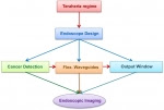
Terahertz (THz) imaging is emerging as a robust platform for a myriad of applications in the fields of security, health, astronomy and material science. The terahertz regime with wavelengths spanning from microns to millimeters is a potentially safe and noninvasive medical imaging modality for detecting cancers. Endoscopic imaging systems provide high flexibility in examining the interior surfaces of an organ or tissue. Researchers have been working on the development of THz endoscopes with photoconductive antennas, which necessarily operate under high voltage, and require at least two channels to measure the reflected signal from the specimen. This manuscript provides the design and imperative steps involved in the development of a single-channel terahertz endoscopic system. The continuous-wave terahertz imaging system utilizes a single flexible terahertz waveguide channel to transmit and collect the back reflected intrinsic terahertz signal from the sample and is capable of operation in both transmission and reflection modalities. To determine the feasibility of using a terahertz endoscope for cancer detection, the co- and cross-polarized terahertz remittance from human colonic tissue specimens were collected at 584 GHz frequency. The two dimensional terahertz images obtained using polarization specific detection exhibited intrinsic contrast between cancerous and normal regions of fresh colorectal tissue. The level of contrast observed using endoscopic imaging correlates well with the contrast levels observed in the free space ex vivo terahertz reflectance studies of human colonic tissue. The prototype device developed in this study represents a significant step towards clinical endoscopic application of THz technology for in vivo colon cancer screening.

Monday, April 11, 2016

OT- SpectroscopyNOW- Reflecting on the prostate: Cancer margins


http://www.spectroscopynow.com/uv/details/highlight/14de3346bad/Last-Months-Most-Accessed-Feature-Reflecting-on-the-prostate-Cancer-margins.html

Reflecting on prostate cancer

Light reflectance spectroscopy can be used to differentiate between malignant and benign prostatic tissue with 85 percent accuracy, according to US researchers. The finding might be used in real-time tissue analysis to guide the surgeon during prostate cancer. Photo UTSouthwestern
Light reflectance spectroscopy, which measure backscattered light, can be used to differentiate between malignant and benign prostatic tissue with 85 percent accuracy, according to US researchers. The finding might be used in real-time tissue analysis to guide the surgeon during prostate cancer.
Urologist Jeffrey Cadeddu of the University of Texas Southwestern Medical Center and colleagues, Aaron Lay, Payal Kapur and Claus Roehrborn, explain that the surgical removal of all cancerous tissue and the sparing of the surrounding healthy tissue during a prostatectomy is not always a completely successful procedure. The removal of healthy tissue can cause problems but leaving behind malignant cells can lead to the recurrence of the prostate cancer.
A real-time approach to guide the surgeon's knife more precisely than ever before could improve the outcome for thousands of men undergoing treatment each year. Radical surgery to remove the prostate gland is often called for, but traditional techniques to assess how much surrounding tissue needs to be removed concurrently are time consuming and do not have proven clinical utility, although cutting too shallow can leave behind problematic cells, "positive surgical margins."

Minimal invasion

"We used a novel light reflectance spectroscopy probe to evaluate surgical margins on radical prostatectomy tissue specimens and correlated the findings with pathological examination," explains Cadeddu; he and his team published details in The Journal of Urology recently. Follow-up studies will be needed to confirm how well the spectroscopic technique might work in the operating theatre.
The team enrolled men with intermediate to high-risk disease requiring radical prostatectomy and examined the prostate tissue following removal with spectroscopic technique, focusing specifically on suspicious malignant and benign prostate margins. Each sample was analyzed and correlated with pathological samples, which were analysed after surgery. In total light reflectance spectroscopy was carried out on 17 prostate gland specimens of which 11 were proven positive histologically; 22 negative surgical margins were measured. The team found that their optical probe could predict positive surgical margins with 85 percent sensitivity, 86 percent specificity, and 86 percent accuracy.

Survival improver?

"This study highlights one of a growing number of technology platforms that aim to improve the outcomes of cancer surgery," explains Cadeddu, an expert in minimally invasive urological surgical techniques. "Further study is required to determine whether such analysis may be used in real time to improve surgical decision-making and decrease the amount of tissue surgeons need to remove."
Prostate cancer is the most common form of the disease in men, second only to skin cancer. It is the second biggest cancer killer in men after lung cancer with more than one in ten affected dying of the disease. "Our next step is to expand to more patients and develop a larger experience to further improve accuracy of technology," Cadeddu told SpectroscopyNOW. "Ultimately, we hope to see the technology commercialize