Showing posts with label biological materials. Show all posts

Friday, August 14, 2020

New phase of nanoconfined water discovered

https://www.nanowerk.com/nanotechnology-news2/newsid=55908.php
(Nanowerk News) Researchers at MIPT Laboratory of Terahertz Spectroscopy together with their Russian and international colleagues discovered a new phase of nanoconfined water; separate water molecules that are confined within nanocavities formed by ions of cordierite crystal lattice.
The first reliable experimental observation of a phase transition in a network of dipole-dipole coupled water molecules is, in and of itself, an important fundamental breakthrough. But apart from that, the discovered phenomenon can also find practical applications in ferroelectrics, artificial quantum systems, and biocompatible nanoelectronics.
The study was a joint effort of MIPT scientists and researchers from Shubnikov Institute of Crystallography, A. M. Prokhorov General Physics Institute of RAS, Skoltech, Sobolev Institute of Geology and Mineralogy, and Novosibirsk State University, as well as their colleagues from Germany (Stuttgart University), the Czech Republic (Prague Institute of Physics), and Japan (University of Tokyo). The results of the study have been reported in Nature Communications ("Dielectric ordering of water molecules arranged in a dipolar lattice").
“We are searching for new phases of electric dipole lattice, i. e. an ensemble of interacting point electric dipoles,” explained Mikhail Belyanchikov, one of the study’s initiators and a junior researcher at MIPT Laboratory of Terahertz Spectroscopy. “A great number of different magnetic dipole phases have been discovered but the research of material phases related not to magnetic but rather to point electric dipoles is still in its early stages. Moreover, electric dipole lattices are a type of ferroelectrics that may have promising microelectronic applications.”


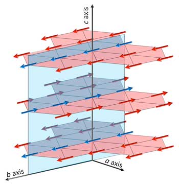
Schematic illustration of ordered state of the electric dipole lattice of polar water molecules inside a cordierite crystal
Schematic illustration of ordered state of the electric dipole lattice of polar water molecules inside a cordierite crystal. Dipole moments are indicated by arrows. The ordered state is manifested through co-existence of ferroelectric (red ab-planes) and antiferroelectric (blue bc-plane) orders. Ferroelectric planes alternate antiferroelectrically along the crystal’s c-axis. (Image courtesy of the researchers)
It is known that to experimentally realize a lattice of point electric dipoles is a challenging task. Usually physicists use the so-called interferometric optical lattice — a periodic structure of fields that is created as the result of laser beams interference. Ultracold atoms of materials to be studied are placed into the lattice points.
But researchers at MIPT Laboratory of Terahertz Spectroscopy found a more efficient way. They place separate water molecules that possess a rather high electric dipole moment into a so-called dielectric matrix, in this case, a zeolite crystal lattice with periodically distributed nanoscale voids formed by lattice ions. One then gets an easily handled sample (a crystal) with practically free water molecules trapped (during crystal growth) in these voids — the so-called nanoconfined water. This sample can be studied in a wide range of temperatures including room temperature and in different environments (electric fields, pressure, etc.).
The key result of the study however was achieved at rather low temperature of 3 K (–270 °C). The studied electric dipole lattice of polar water molecules was based on a cordierite crystal — a member of the zeolites family. The researchers observed an order-disorder ferroelectric phase transition in a three-dimensional nanoconfined water molecular network at the temperature of 3 K.
“Previously, we had studied similar nanoconfined water molecules located within a matrix of beryl, a crystal that possesses the structure very similar to that of cordierite. We did not register ordering of molecular dipoles in this system even at 0.3 K, the lowest temperature we were able to achieve. The reason may be the relatively high symmetry (hexagonal) of beryl crystal lattice and the quantum-mechanical phenomena that govern properties of water at such low temperatures,” noted Mikhail Belyanchikov. “At the same time, it is cordierite’s somewhat lower (orthorhombic) crystalline symmetry that triggered the phase transition in an array of water molecules hosted by its crystal lattice.”
To analyze and interpret experimental findings, researchers employed computer modeling. Monte Carlo simulation and other mathematical methods were used for numerical solution of the extremely complex multiparticle Schrödinger equation describing the electric dipole system of interacting polar water molecules.
Computer modeling helped visualize the ordered phase at microscopic — or rather nanoscopic — scale. And yet again, the scientists were taken by surprise as this phase turned out to be rather unusual. It is manifested as co-existence of ferroelectric and antiferroelectric orderings of water dipole moments. It can be visualized as a stack of alternating sheets of co-aligned dipoles where dipoles in every two adjacent sheets are oriented antiparallel (see the figure above).
The simulations also showed that the structure of ordered water dipoles (arrows in the figure) can be even more complex. This happens when water molecules only fill some of the crystal’s cavities. In that case, dipole arrows in the sheets group in separate domains.
“Not only does studying nanoconfined water molecules have a fundamental importance for the field of electro-dipolar lattices but it also contributes to deeper understanding of natural phenomena and may even potentially enable construction of biocompatible nanoelectronic devices. This is a rapidly developing field that promises new and extremely efficient electronics based on biological materials,” comments Boris Gorshunov, who heads MIPT Laboratory of Terahertz Spectroscopy.
Source: Moscow Institute of Physics and Technology

Tuesday, July 2, 2019

Kirigami can spin terahertz rays in real time to peer into biological tissue


https://news.umich.edu/kirigami-can-spin-terahertz-rays-in-real-time-to-peer-into-biological-tissue/

ANN ARBOR—With a light-spinning device inspired by the Japanese art of paper cutting, University of Michigan researchers have detected microscopic twists in the internal structure of plant and animal tissue without harmful X-rays.
The approach is the first that can fully rotate terahertz radiation in real time, and it could open new dimensions in medical imaging, encrypted communications and cosmology. The researchers are most interested in using terahertz rays to identify biological tissues through the twists in their structures—their “chirality.” A tissue’s chirality affects how much it absorbs twisted radiation.
Terahertz radiation is the band of electromagnetic waves that runs from infrared radiation down to the range of the “millimeter scanners” that peer through your clothing at airports. It can travel about a quarter of an inch into the body, but unlike X-rays, it’s non-ionizing—meaning it doesn’t free up potentially damaging electrical charges in the body. 
“Our bodies have a lot of twisted structures that are close enough to the surface for terahertz photons to penetrate: vessels, ligaments, muscle fibers, molecules and even some helical bacteria,” said Nicholas Kotov, the Joseph B. and Florence V. Cejka Professor of Engineering and a corresponding author on the study in Nature Materials.
He believes it may be possible to gain medically relevant information about the working behaviors of these tissues using terahertz imaging. However, as with X-rays, it is difficult to tell the difference between soft tissues in terahertz scans. 
With an eye to exploring how chirality may help distinguish tissues, the team gathered everyday biological materials to look for differences in the absorption of clockwise- or counter-clockwise-rotating radiation in the terahertz spectrum. They studied a maple leaf, a dandelion flower, pork fat and the wing case of an iridescent beetle. While the leaf and fat showed no difference in absorption of clockwise or counter-clockwise radiation, the flower and wing case preferentially absorbed the one over the other, revealing microscopic twists in their structures.
This technique, called circular dichroism spectroscopy, was impractical in the terahertz range until now. Other parts of the electromagnetic spectrum, such as visible light, can be twisted with natural crystals, but the twisting power was limited for terahertz radiation or else it couldn’t be done in real time. 
The new device is a deceptively simple—essentially a plastic ribbon, printed with a gold herringbone pattern and sliced with staggered rows of tiny cuts. The incisions are influenced by the Japanese art of kirigami, which uses arrangements of cuts to create 3D structures from paper.
When the ribbon is stretched, the cuts open up and the slices of ribbon twist. The gold lines then guide the radiation, twisting it in turn. In radiation, twisting is called “circular polarization,” which is the same optical phenomenon that is used in liquid crystal displays (LCDs). 
“We all might have an experience of playing with paper crafting when we were young, but there were no design rules for a 3D chiral optical devices built using only folding and cutting. So, we started from scratch and tested many models through both simulations and experiments,” said Wonjin Choi, a Ph.D. student in materials science and engineering and co-first author on the study.
The team proposes the same design could be scaled for other types of radiation as well, with larger patterns interacting with microwaves or radio waves, or shrinking the pattern down to manipulate infrared light.
Because spinning terahertz light wasn’t widely studied, one of the team’s challenges was figuring out how to see whether the kirigami device worked at all.
“The conventional ways of measuring terahertz radiation are limited to how much energy is lost as it travels through a sample, which is not enough for our case,” said Gong Cheng, a Ph.D. student in physics at U-M and co-first author.
By stacking linear polarizers, rotated with respect to one another, in the path of the beam, they could make measurements to reveal the circular polarization.
In addition to imaging living tissues, terahertz circular dichroism spectroscopy could also aid the development of new medicines based on large biological molecules such as proteins and antibodies. 
Choi anticipates that an early application could be to encrypt and decrypt communications on the terahertz spectrum. And if these kirigami devices were flown on satellites to measure the twist in the terahertz spectrum of the universe’s background radiation, it could tell us more about the earliest stars.
The study was supported by the Defense Advanced Research Projects Agency and the Department of Defense’s Vannevar Bush Fellowship. The chiral kirigami modulator was built in the Lurie Nanofabrication Facility.
Ted Norris, the Gerard A. Mourou Collegiate Professor of Electrical Engineering and Computer Science, is also a corresponding author on the study. Kotov is also a professor of chemical engineering, materials science and engineering and macromolecular science and engineering.