Showing posts with label Hwayeong Cheon. Show all posts

Thursday, June 25, 2020

Abstract-Transformation of terahertz vibrational modes of cytosine under hydration


Donggun Lee, Hwayeong Cheon, Seo-Yeon Jeong, Joo-Hiuk Son
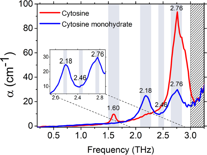
figure1
https://www.nature.com/articles/s41598-020-67179-z

Cytosine and cytosine monohydrate are representative biomolecules for investigating the effect of hydrogen bonds in deoxyribonucleic acid. To better understand intermolecular interactions, such as hydrogen bonds, between nucleobases it is necessary to identify the low-frequency vibrational modes associated with intermolecular interactions and crystalline structures. In this study, we investigated the characteristic low-frequency vibrational modes of cytosine and cytosine monohydrate using terahertz time-domain spectroscopy (THz-TDS). The crystal geometry was obtained by the powder X-ray diffraction technique. The optimized atomic positions and the normal modes in the terahertz region were calculated using density functional theory (DFT), which agreed well with the experimental results. We found that overall terahertz absorption peaks of cytosine and cytosine monohydrate consist of collective vibrations mixed with intermolecular and intramolecular vibrations in mode character analysis, and that the most intense peaks of both samples involve remarkable intermolecular translational vibration. These results indicate that THz-TDS combined with DFT calculations including mode character analysis can be an effective method for understanding how water molecules contribute to the characteristics of the low-frequency vibrational modes by intermolecular vibrations with hydrogen bonding in biological and biomedical applications.

Saturday, February 1, 2020

Abstract-Determining terahertz resonant peaks of biomolecules in aqueous environment


Seo-Yeon Jeong, Hwayeong Cheon, Donggun Lee, and Joo-Hiuk Son


Sample holder for maintaining constant temperature of the sample. (a) Structure of sample holder. Inside the holder, a pair of thermoelectric cooling devices were in contact with the copper plate. The thermoelectric devices were cooled by the water cooler. The copper plate had a hole in the middle, and the quartz window was mounted inside the hole. (b) Process of filling liquid sample inside the container. The z-cut quartz window was fixed with a cylinder ring. The 300-µm copper spacer was placed on the z-quartz window. The liquid sample was dropped in the middle of spacer and covered with a Teflon window for 5 min. The Teflon window was removed after the sample was fully frozen, and the experiments were performed at an equilibrium temperature of 250 K.

https://www.osapublishing.org/oe/abstract.cfm?uri=oe-28-3-3854

The resonant peaks of biomolecules provide information on the molecules’ physical and chemical properties. Although many biomolecules have resonant peaks in the terahertz region, it is difficult to observe their specific signals in aqueous environments. Hence, this paper proposes a method for determining these peaks. We found the specific resonant peaks of a modified nucleoside, 5-methlycytidine and modified HEK293T DNA in an aqueous solution through baseline correction. We evaluated the consistency of various fitting functions used for determining the peaks with various parameters. We separated two resonance peaks of 5-methlycytidine at 1.59 and 1.97 THz and for artificially methylated HEK293T DNA at 1.64 and 2.0 THz.
© 2020 Optical Society of America under the terms of the OSA Open Access Publishing Agreement

Sunday, September 8, 2019

Abstract-Effective demethylation of melanoma cells using terahertz radiation


Hwayeong Cheon, Hee-Jin Yang, Moran Choi, and Joo-Hiuk Son

 Schematic showing THz demethylation using resonant THz radiation system. High-power THz radiation was generated using a regenerative amplifier and LiNbO3 crystal. The THz filter limited the THz bandwidth to around the resonance frequency of the methyl-DNA bonds.


https://www.osapublishing.org/boe/abstract.cfm?uri=boe-10-10-4931

Terahertz (THz) demethylation is a photomedical technique applied to dissociate methyl-DNA bonds and reduce global DNA methylation using resonant THz radiation. We evaluated the performance of THz demethylation and investigated the DNA damage caused by THz irradiation. The demethylation rate in M-293T DNA increased linearly with the irradiation power up to 48%. The degree of demethylation increased with exposure to THz radiation, saturating after 10 min. Although THz demethylation occurred globally, most of the demethylation occurred within the partial genes in the CpG islands. Subsequently, we performed THz demethylation of melanoma cells. The degree of methylation in the melanoma cell pellets decreased by approximately 10–15%, inducing ∼5–8 abasic sites per 105 bp; this was considerably less than the damaged DNA irradiated by the high-power infrared laser beam used for generating THz pulses. These results provide initial data for THz demethylation and demonstrate the applicability of this technique in advanced cancer cell research. THz demethylation has the potential to develop into a therapeutic procedure for cancer, similar to that involving chemical demethylating agents.
© 2019 Optical Society of America under the terms of the OSA Open Access Publishing Agreement

Wednesday, May 1, 2019

Abstract-Detection and manipulation of methylation in blood cancer DNA using terahertz radiation


Hwayeong Cheon, Jin Ho Paik, Moran Choi, Hee-Jin Yang,  Joo-Hiuk Son


https://www.nature.com/articles/s41598-019-42855-x

DNA methylation is a pivotal epigenetic modification of DNA that regulates gene expression. Abnormal regulation of gene expression is closely related to carcinogenesis, which is why the assessment of DNA methylation is a key factor in cancer research. Terahertz radiation may play an important role in active demethylation for cancer therapy because the characteristic frequency of the methylated DNA exists in the terahertz region. Here, we present a novel technique for the detection and manipulation of DNA methylation using terahertz radiation in blood cancer cell lines. We observed the degree of DNA methylation in blood cancer at the characteristic resonance of approximately 1.7 THz using terahertz time-domain spectroscopy. The terahertz results were cross-checked with global DNA methylation quantification using an enzyme-linked immunosorbent assay. We also achieved the demethylation of cancer DNA using high-power terahertz radiation at the 1.7-THz resonance. The demethylation degrees ranged from 10% to 70%, depending on the type of cancer cell line. Our results show the detection of DNA methylation based on the terahertz molecular resonance and the manipulation of global DNA methylation using high-power terahertz radiation. Terahertz radiation may have potential applications as an epigenetic inhibitor in cancer treatment, by virtue of its ability to induce DNA demethylation, similarly to decitabine.

Monday, November 12, 2018

Abstract-Toward Cancer Treatment Using Terahertz Radiation: Demethylation of Cancer DNA


Joo-Hiuk Son, Hwayeong Cheon

https://ieeexplore.ieee.org/document/8510290

Carcinogenesis involves DNA methylation which is a primary alteration in DNA in the development of cancer occurring before genetic mutation. Because the abnormal DNA methylation is found in most of cancer cells, the detection and manipulation of DNA methylation using terahertz radiation can be a novel pioneering method in cancer study. The DNA methylation has been directly observed by terahertz spectroscopy at around 1.65 THz and this epigenetic chemical change could be manipulated to the state of demethylation using a high-power terahertz radiation. Demethylation of cancer DNA is a key problem in epigenetic cancer therapy and our results may lead to the treatment of cancer in early stage.

Wednesday, May 17, 2017

Abstract-Toward clinical cancer imaging using terahertz spectroscopy


Hwayeong Cheon ; Hee-Jin Yang ;  Joo-Hiuk Son

http://ieeexplore.ieee.org/document/7929316/

Cancer imaging using terahertz (THz) electromagnetic waves has the potential to overcome the drawbacks of existing cancer imaging techniques because of the unique properties of THz radiation. It is non-ionizing, highly sensitive to water molecules, and suitable for the observation of many biomolecular characteristics based on low-energy vibrational modes. Consequently, it is advantageous to use THz cancer imaging for detection, especially of superficial carcinomas in soft tissues. However, there are three primary challenges facing this type of cancer imaging that must be addressed before it can be applied medically: the limited penetration depth in hydrated tissues, the difficulty of obtaining molecular resonance fingerprints of cancers, and the low image contrast between tissues. These challenges can be overcome by applying several state-of-the-art techniques; the penetration depth has been enhanced sufficiently to observe cancer lesions deep inside tissues by using freezing and penetration-enhancing agents: the biochemical modification of DNA can be utilized to track the resonance fingerprints of carcinogenesis at the genomic DNA level; and nanoparticles can increase the THz imaging contrast if they are employed similarly to how they are used in magnetic resonance imaging. These solutions are important to enable THz cancer imaging to be performed in clinical settings.

Thursday, January 26, 2017

Abstract-Terahertz imaging of metastatic lymph nodes using spectroscopic integration technique


Jae Yeon Park, Hyuck Jae Choi, Hwayeong Cheon, Seong Whi Cho, Seungkoo Lee, and Joo-Hiuk Son

https://www.osapublishing.org/boe/abstract.cfm?uri=boe-8-2-1122


Terahertz (THz) imaging was used to differentiate the metastatic states of frozen lymph nodes (LNs) by using spectroscopic integration technique (SIT). The metastatic states were classified into three groups: healthy LNs, completely metastatic LNs, and partially metastatic LNs, which were obtained from three mice without infection and six mice infected with murine melanoma cells for 30 days and 15 days, respectively. Under histological examination, the healthy LNs and completely metastatic LNs were found to have a homogeneous cellular structure but the partially metastatic LNs had interfaces of the melanoma and healthy tissue. THz signals between the experimental groups were not distinguished at room temperature due to high attenuation by water in the tissues. However, a signal gap between the healthy and completely metastatic LNs was detected at freezing temperature. The signal gap could be enhanced by using SIT that is a signal processing method dichotomizing the signal difference between the healthy cells and melanoma cells with their normalized spectral integration. This technique clearly imaged the interfaces in the partially metastatic LNs, which could not be achieved by existing methods using a peak point or spectral value. The image resolution was high enough to recognize a metastatic area of about 0.7 mm size in the partially metastatic LNs. Therefore, this pilot study demonstrated that THz imaging of the frozen specimen using SIT can be used to diagnose the metastatic state of LNs for clinical application.
© 2017 Optical Society of America
Full Article  |  PDF Article

Tuesday, November 15, 2016

Abstract-Terahertz molecular resonance of cancer DNA

http://www.nature.com/articles/srep37103

Carcinogenesis involves the chemical and structural alteration of biomolecules in cells. Aberrant methylation of DNA is a well-known carcinogenic mechanism and a common chemical modification of DNA. Terahertz waves can directly observe changes in DNA because the characteristic energies lie in the same frequency region. In addition, terahertz energy levels are not high enough to damage DNA by ionization. Here, we present terahertz molecular resonance fingerprints of DNA methylation in cancer DNA. Methylated cytidine, a nucleoside, has terahertz characteristic energies that give rise to the molecular resonance of methylation in DNA. Molecular resonance is monitored in aqueous solutions of genomic DNA from cancer cell lines using a terahertz time-domain spectroscopic technique. Resonance signals can be quantified to identify the types of cancer cells with a certain degree of DNA methylation. These measurements reveal the existence of molecular resonance fingerprints of cancer DNAs in the terahertz region, which can be utilized for the early diagnosis of cancer cells at the molecular level.