Showing posts with label Hans Bechtel. Show all posts

Thursday, May 8, 2014

Berkeley Lab Develops Nanoscope to Probe Chemistry on the Molecular Scale


http://newscenter.lbl.gov/news-releases/2014/05/07/berkeley-lab-develops-nanoscope-to-probe-chemistry-on-the-molecular-scale/?fb_action_ids=10202078686957296&fb_action_types=og.likes

New technique uses infrared synchrotron light and atomic force microscopy to study batteries, cell membranes, stardust, and other complex systems on the nanoscale

Kate Greene 510-486-4404  kgreene@lbl.gov
 0 

 
News Release


For years, scientists have had an itch they couldn’t scratch. Even with the best microscopes and spectrometers, it’s been difficult to study and identify molecules at the so-called mesoscale, a region of matter that ranges from 10 to 1000 nanometers in size. Now, with the help of broadband infrared light from the Advanced Light Source (ALS) synchrotron at the U.S. Department of Energy’s Lawrence Berkeley National Laboratory (Berkeley Lab), researchers have developed a broadband imaging technique that looks inside this realm with unprecedented sensitivity and range.
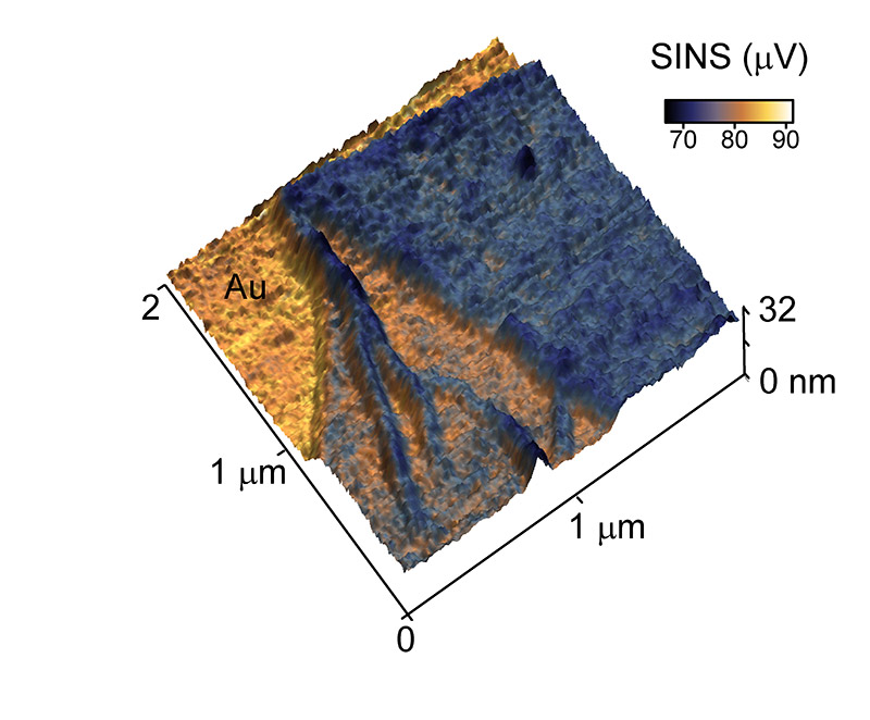
This peptoid nanosheet, produced by Gloria Olivier and Ron Zuckerman at Berkeley Lab, is less than 8 nanometers thick at points. SINS makes it possible to acquire spectroscopic images of these ultra-thin nanosheets for the first time.
By combining atomic force microscopy with infrared synchrotron light, researchers from Berkeley Lab and the University of Colorado have improved the spatial resolution of infrared spectroscopy by orders of magnitude, while simultaneously covering its full spectroscopic range, enabling the investigation of variety of nanoscale, mesoscale, and surface phenomena that were previously difficult to study.
The new technique, called Synchrotron Infrared Nano-Spectroscopy or SINS, will enable in-depth study of complex molecular systems, including liquid batteries, living cells, novel electronic materials and stardust.
“The big thing is that we’re getting full broadband infrared spectroscopy at 100 to 1000 times smaller scale,” says Hans Bechtel, principal scientific engineering associate at Berkeley Lab. “This is not an incremental achievement. It’s really revolutionary.”
In a Proceedings of the National Academy of Sciences paper published May 6 onlinetitled “Ultra-broadband infrared nano-spectroscopic imaging,” Berkeley Lab’s Bechtel and Michael Martin, a Berkeley Lab staff scientist, and colleagues from Markus Raschke’s group at the University of Colorado at Boulder describe SINS. They demonstrate the nanoscope’s ability to capture broadband spectroscopic data over a variety of samples, including a semiconductor-insulator system, a mollusk shell, proteins, and a peptoid nanosheet. Martin says these demonstrations just “scratch the surface” of the potential of the new technique.
Synchronizing Scopes
SINS combines two pre-existing infrared technologies: a newer technique called infrared scattering-scanning near-field optical microscopy (IR s-SNOM) and an old laboratory standby, known even to college chemistry students, called Fourier Transform Infrared Spectroscopy (FTIR). A clever melding of these two tools, combined with the intense infrared light of the synchrotron at Berkeley Lab gives the researchers the ability to identify clusters of molecules sized as small as 20 to 40 nanometers.
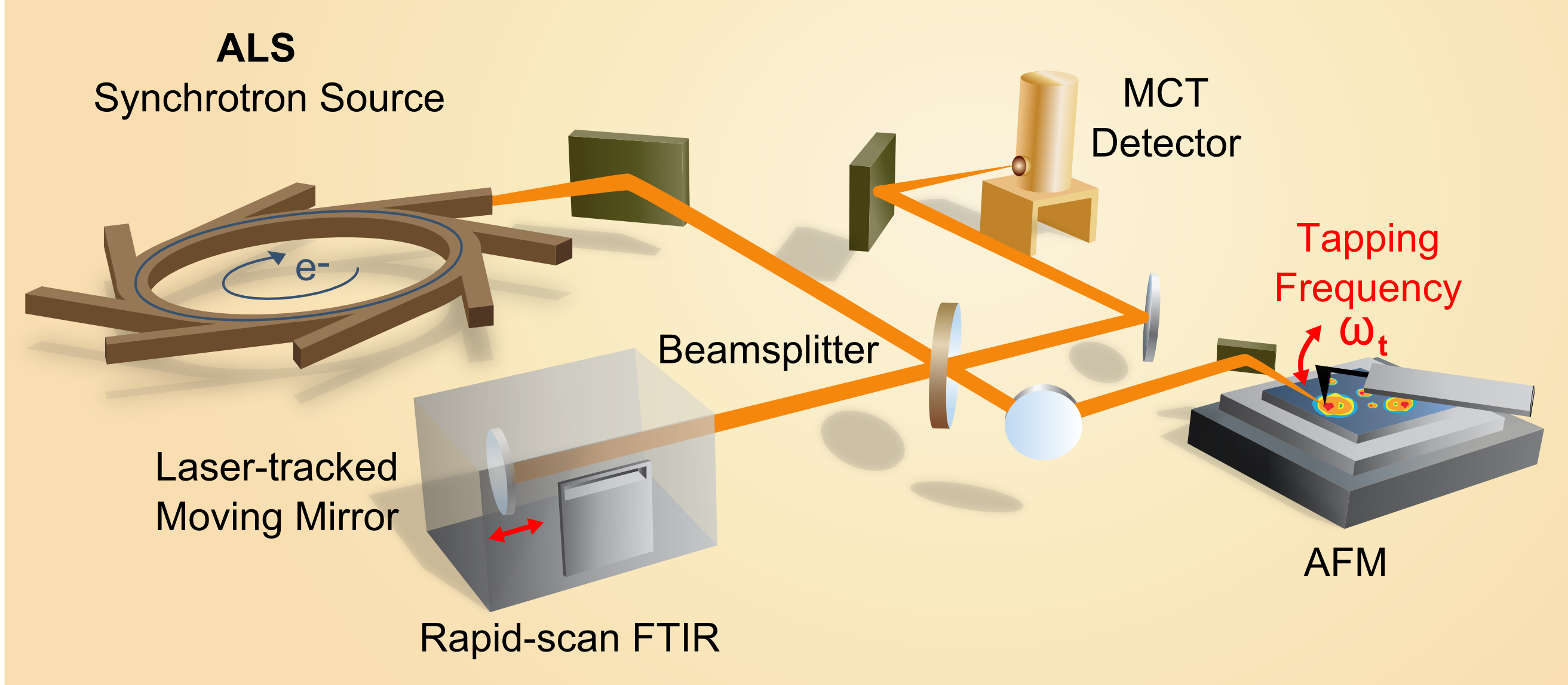
Experimental setup for SINS that includes the synchrotron light source, an atomic force microscope, a rapid-scan Fourier transform infrared spectrometer, a beamsplitter, mirrors and a detector.
The new approach overcomes long-standing barriers with pre-existing microscopy techniques that often involve demanding technical and sample preparation requirements. Infrared spectroscopy uses low-energy light, is minimally invasive, and is applicable under ambient conditions, making it an excellent tool for chemical and molecular identifications in systems that are static as well as those that are living and dynamic. The technique works by shining low-energy infrared light onto a molecular sample. Molecules can be thought of as systems of balls (atoms) and springs (bonds between atoms) that vibrate with characteristic wiggles; they absorb infrared radiation at frequencies that correspond to their natural vibrating modes. The output from this absorption is a spectrum, often called a fingerprint, which shows distinctive peaks and dips, depending on the bonds and atoms present in the sample.
But infrared spectroscopy has its challenges too. While it works well for bulk samples, traditional infrared spectroscopy can’t resolve molecular composition below about 2000 nanometers. The major hurdle is the diffraction limit of light, which is the fundamental barrier that determines the smallest focus spot of light and is particularly troublesome for the large wavelengths of infrared light. In recent years, though, the diffraction limit has been overcome by a technique called scattering-scanning near-field optical microscopy, or s-SNOM, which involves shining light onto a metallic tip. The tip acts as an antenna for the light, directing it to a tiny region at its apex just tens of nanometers wide.
This trick is what’s used in IR s-SNOM, where infrared light is coupled to a metallic tip. The challenge with IR s-SNOM, however, is that researchers have been relying on infrared light produced by lasers. Lasers emit a large number of photons needed for the technique, but because they operate in a narrow wavelength band, they can only probe a narrow range of molecular vibrations. In other words, laser light simply can’t give you the flexibility to explore a spectrum of mixed molecules.
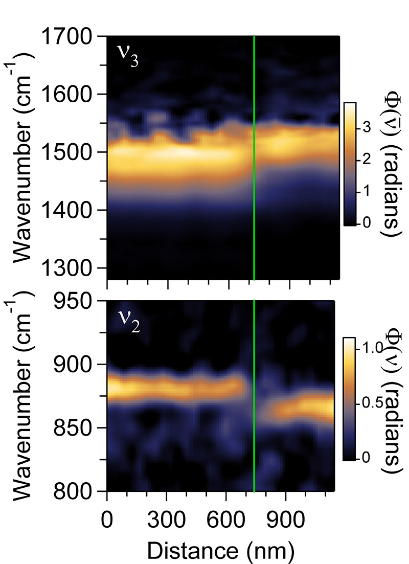
A spectral-linescan of a blue mussel shell, which transitions from calcite to aragonite, illustrates the spatial resolution and spectroscopic range capabilities of the SINS technique. The image shows two simultaneously acquired vibrational modes across the transition region.
Bechtel, Martin and Raschke’s team saw the opportunity to use Berkeley Lab’s ALS to overcome the laser limitation. The lab’s synchrotron produces broadband infrared light with a high-photon count that can be focused to the diffraction limit. The researchers coupled the synchrotron light to a metallic tip with an apex of about 20 nanometers, focusing the infrared beam onto the samples. The resulting spectrum is analyzed with a modified FTIR instrument.
“This is actually one of very few examples where synchrotron light has been coupled to scanning probe microscopy,” says Raschke. “Moreover, the implementation of the technique at the synchrotron brings chemical nano-spectroscopy and -imaging out of the lab of a few laser science experts and makes it available for a broader scientific community at a user facility.”
From mollusks to moon rocks
The team demonstrated the technique by confirming the spectroscopic signature of silicon dioxide on silicon and by illustrating the sharp chemical transition that occurs within the shells of the blue mussel (M. edulis). Additionally, the researchers looked at proteins and a peptoid nanosheet, an engineered, ultra-thin film of proteins with medical and pharmacological applications.
Martin is excited for the potential of SINS, which is available for researchers from any institution to use. In particular he’s interested in a closer look at battery systems, with the hope that understanding battery chemistry on the mesoscale could provide insight into better performance. Further out, he expects SINS to be useful for a range of biochemistry as well. “This hints at a dream I’ve had in my mind, to look at the surface of a cell, inside the bi-layer membrane, the channels, and receptors,” says Martin. “If we could put a SINS tip on a living cell, we could watch biochemistry happen in real time.”
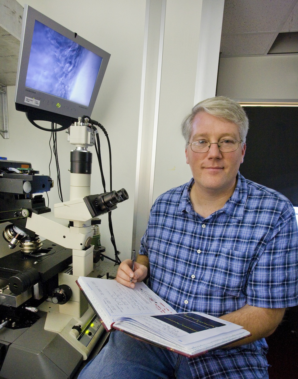
Berkeley Lab's Michael Martin
Bechtel, for his part, is intrigued by the possibility of using SINS for the study of lunar rocks, meteorites and stardust. These extraterrestrial materials have a molecular diversity that is difficult to resolve on the nanoscale, particularly in a non-destructive manner for these rare samples. A better understanding of the makeup of moon rocks and dust from space could provide clues to the formation of the planets and solar system.
Raschke is using the technique to study the processes that limit the performance of organic solar cells. He is looking to further improve the flexibility of the technique such that it can be applied under variable and controlled atmospheric and low-temperature conditions. Among other tweaks, he plans to increase the sensitivity of the technique with the ultimate goal of performing singe-molecule chemical spectroscopy.
This research was supported by the DOE Office of Science.
###
Lawrence Berkeley National Laboratory addresses the world’s most urgent scientific challenges by advancing sustainable energy, protecting human health, creating new materials, and revealing the origin and fate of the universe. Founded in 1931, Berkeley Lab’s scientific expertise has been recognized with 13 Nobel prizes. The University of California manages Berkeley Lab for the U.S. Department of Energy’s Office of Science. For more, visit www.lbl.gov.
The Advanced Light Source is a third-generation synchrotron light source producing light in the x-ray region of the spectrum that is a billion times brighter than the sun. A DOE national user facility, the ALS attracts scientists from around the world and supports its users in doing outstanding science in a safe environment. For more information visit www-als.lbl.gov.
DOE’s Office of Science is the single largest supporter of basic research in the physical sciences in the United States and is working to address some of the most pressing challenges of our time. For more information, please visit the Office of Science website atscience.energy.gov/.

Wednesday, May 2, 2012

Molecular Spectroscopy Tracks Living Mammalian Cells in Real Time as They Differentiate

(Berkeley Lab scientists observed phosphorylation in living PC12 cells stimulated by nerve growth factor as they differentiated and sent out neuron-like neurites. The researchers imaged individual cells and simultaneously obtained absorption spectra using synchrotron radiation from the Advanced Light Source. Cells not stimulated with nerve growth factor did not differentiate and showed different infrared absorption spectra)   http://newscenter.lbl.gov/feature-stories/2012/04/30/phosphorylation/

Berkeley Lab scientists demonstrate the promise of synchrotron infrared spectroscopy of living cells for medical applications

April 30, 2012

Knowing how a living cell works means knowing how the chemistry inside the cell changes as the functions of the cell change. Protein phosphorylation, for example, controls everything from cell proliferation to differentiation to metabolism to signaling, and even programmed cell death (apoptosis), in cells from bacteria to humans. It’s a chemical process that has long been intensively studied, not least in hopes of treating or eliminating a wide range of diseases. But until now the close-up view – watching phosphorylation work at the molecular level as individual cells change over time – has been impossible without damaging the cells or interfering with the very processes that are being examined.
“To look into phosphorylation, researchers have labeled specific phosphorylated proteins with antibodies that carry fluorescent dyes,” says Hoi-Ying Holman of the U.S. Department of Energy’s Lawrence Berkeley National Laboratory (Berkeley Lab). “That gives you a great image, but you have to know exactly what to label before you can even begin.”
Holman and her coworkers worked with colleagues from the San Diego and Berkeley campuses of the University of California to develop a new technique for monitoring protein phosphorylation inside single living cells, tracking them over a week’s time as they underwent a series of major changes.
“Now we can follow cellular chemical changes without preconceived notions of what they might be,” says Holman, a pioneer in infrared (IR) studies of living cells who is director of the Berkeley Synchrotron Infrared Structural Biology program at Berkeley Lab’s Advanced Light Source (ALS) and head of the Chemical Ecology Research group in the Earth Sciences Division . “We’ve monitored unlabeled living cells by studying the nonperturbing absorption of a wide spectrum of bright synchrotron infrared radiation from the ALS.”
The researchers report their results in the American Chemical Society journal Analytical Chemistry.
Phosphorylation fundamentals
Phosphorylating enzymes add one or more phosphate groups to three amino-acid residues common in proteins – serine, threonine, or tyrosine – which activates the proteins; removing the phosphate reverses the process. The research goal is to learn exactly when proteins such as enzymes and receptors are switched on and off by phosphorylation, and which cells within a population are responding to cause specific changes – for example, during differentiation of a progenitor cell into its functional form.
To avoid killing cells or introducing modified proteins or foreign bodies that may alter their behavior, scientists can use a method called Fourier-transform infrared (FTIR) spectromicroscopy; because infrared light has lower photon energy than x-rays, it can peer inside living cells without damaging them. Different components and different states of the cell absorb different wavelengths of the broad infrared spectrum; applying the Fourier-transform algorithm allows signals of all frequencies to be recorded simultaneously, pinpointing when, where, and what chemical changes are occurring.
Most infrared sources are dim, however, so the information from typical IR set-ups is limited in resolution and has a low signal-to-noise ratio. Infrared from the ALS’s synchrotron light source is a hundred to a thousand times brighter.
PC12 cells treated with nerve growth factor underwent a series of changes due to phosphorylation. Beginning at Day 3 they sent out neurites, resembling the growth of nerve cells. Spectromicroscopy at beamline 1.4 of the Advanced Light Source tracked specific local chemical changes in the living cells.
PC12 cells treated with nerve growth factor underwent a series of changes due to phosphorylation. Beginning at Day 3 they sent out neurites, resembling the growth of nerve cells. Spectromicroscopy at beamline 1.4.3 of the Advanced Light Source tracked specific local chemical changes in the living cells.
Previously Holman and her colleagues have used IR beamline 1.4.3, managed by Berkeley Lab’s Michael Martin and Hans Bechtel, to obtain spectra from living organisms in rock, soil, and water. They have monitored ongoing biochemistry within living bacteria adapting to stress, and more recently within individual skin connective tissue cells (fibroblasts) from patients with mitochondrial disorders. (Mitochondria are the cellular organelles commonly known as the “power-plants” of the cell.)
The present study was done with a line of cultured cells called PC12. When nerve growth factor, a small protein, is introduced into a PC12 cell, the cell begins to send out neurites resembling the projections from nerve cell bodies. Although originally derived from a tumor of the rat’s adrenal gland, PC12 has become, rather counterintuitively, a valuable model of how nerve cells differentiate from their unspecialized progenitors.
Berkeley Lab postdoctoral fellow Liang Chen began the current experiments by introducing nerve growth factor to groups of PC12 cells to induce them to differentiate; one group of cells was left untreated as a control. The cells were cultured on gold-coated slides in chambers maintained at body temperature in a humidified environment and supplied with nutrients. Individual cells of a group were positioned under the infrared beam at the beamline 1.4.3 endstation.
FTIR spectra were collected before and after the nerve growth factor was introduced. After stimulation, the spectra were taken first at short intervals, from two to sixty minutes apart. Additional spectra were collected of cells in other groups on the third, fifth, and seventh day of continued stimulation.
The first day’s spectra revealed spikes in phosphorylation activity within minutes after the addition of the nerve growth factor, in concert with changes in the ratios of such important chemical contents of the cell as proteins, carbohydrates, and lipids. Phosphorylation subsequently waned, then picked up again in another burst of activity on Day 3, just as the cells began to extend neurites.
By comparing results with quantum chemistry simulations by Berkeley Lab’s Zhao Hao — predicting what should be observed from first principles — as well as with results from partial studies using other methods, the researchers confirmed the monitoring of phosphorylation phases, their timing, and their target proteins, along with associated changes in other substances in the cell.
In the top panel, different modes of imaging of the same cell show the differences between visible light microscopy and fluorescence imaging, and in the lower panel, the images resulting from Fourier-transform infrared spectromicroscopy.  Infrared absorption at different frequencies pinpoints different cell components at specific locations in the living cell. (Click on image for best resolution.)
In the top panel, different modes of imaging of the same cell show the differences between visible light microscopy and fluorescence imaging, and in the lower panel, the images resulting from Fourier-transform infrared spectromicroscopy. Infrared absorption at different frequencies pinpoints different cell components at specific locations in the living cell. 
A new technique takes off
“This experiment was a proof of the concept,” says Liang Chen. “We demonstrated the dynamics of protein phosphorylation in controlling differentiation in this biological system using synchrotron infrared spectromicroscopy, and we pointed the way to answering the many questions a biologist has to ask about measuring the coordination of specific processes in real time.”
Although in this first experiment the team was not able to follow individual cells continuously, they were able to monitor differentiation in groups of cultured PC12 cells in real time, without labeling or any other invasive procedure. It was the first step in an ambitious inquiry into the real-time biochemistry of living mammalian cells over the long term.
At beamline 1.4.3., with the help of new team members Kevin Loutherback and Rafael Gomez-Sjoberg, the team is designing equipment to maintain mammalian cells in a thin layer of culture media that will keep them healthy yet not interfere with the infrared beam, while automatically monitoring and adjusting temperature, humidity, and nutrient ratios, and removing waste products. This will allow data on individual cells to be gathered continuously throughout the entire phosphorylation process.
Meanwhile the Berkeley Synchrotron Infrared Structural Biology program at ALS beamline 5.4 is building multimodal facilities that will monitor cell development in human cells, bacteria, and plants, within soils, minerals, and other environments, via “hyperspectromicroscopy” – from the ultraviolet through visible light and deep into the infrared. Researchers will be able to choose the frequency window (or combination of windows) best suited to the sample and the conditions – in Holman’s words, “to watch almost everything at once.”
Says Holman, “Many researchers from the medical communities are interested in using the technology, and we are particularly interested in collaborating with university centers and private firms that are seeking a broad view of how promising drugs act within specific cells.”
Some of the projects will target Alzheimer’s disease, macular degeneration of the retina in diabetes, and mitochondrial diseases in children. In addition, specific processes like protein glycation can also be identified. Since different cells and different organisms respond differently, the eventual goal is to develop specific ways to screen the mechanisms of individual medicines.
###
“Synchrotron infrared measurements of protein phosphorylation in living single PC12 cells during neuronal differentiation,” by Chen et al, appears in Analytical Chemistry, online at http://pubs.acs.org/doi/full/10.1021/ac300308x.
The research was performed at the Advanced Light Source and the Berkeley Synchrotron Infrared Structural Biology Program, which are supported by the U.S. Department of Energy Office of Science.
Learn more about infrared beamlines at the Advanced Light Source at infrared.als.lbl.gov/.
The Advanced Light Source is a third-generation synchrotron light source producing light in the x-ray region of the spectrum that is a billion times brighter than the sun. A DOE national user facility, the ALS attracts scientists from around the world and supports its users in doing outstanding science in a safe environment. For more information visit www-als.lbl.gov/.
DOE’s Office of Science is the single largest supporter of basic research in the physical sciences in the United States, and is working to address some of the most pressing challenges of our time. For more information, please visit the Office of Science website at science.energy.gov/.
Lawrence Berkeley National Laboratory addresses the world’s most urgent scientific challenges by advancing sustainable energy, protecting human health, creating new materials, and revealing the origin and fate of the universe. Founded in 1931, Berkeley Lab’s scientific expertise has been recognized with 13 Nobel prizes. The University of California manages Berkeley Lab for the U.S. Department of Energy’s Office of Science. For more, visit www.lbl.gov.

Sunday, September 4, 2011

Tunable graphene device demonstrated: First tool in kit for putting terahertz light to work


The graphene microribbon array can be tuned in three ways. Varying the width of the ribbons changes plasmon resonant frequency and absorbs corresponding frequencies of terahertz light. Plasmon response is much stronger when there is a dense concentration of charge carriers (electrons or holes), controlled by varying the top gate voltage. Finally, light polarized perpendicularly to the ribbons is strongly absorbed at the plasmon resonant frequency, while parallel polarization shows no such response. Credit: Lawrence Berkeley National Laboratory
Long-wavelength terahertz light is invisible – it's at the farthest end of the far infrared – but it's useful for everything from detecting explosives at the airport to designing drugs to diagnosing skin cancer. Now, for the first time, scientists at the U.S. Department of Energy's Lawrence Berkeley National Laboratory (Berkeley Lab) and the University of California at Berkeley have demonstrated a microscale device made of graphene – the remarkable form of carbon that's only one atom thick – whose strong response to light at terahertz frequencies can be tuned with exquisite precision.

The heart of our device is an array made of  ribbons only millionths of a meter wide," says Feng Wang of Berkeley Lab's Materials Sciences Division, who is also an assistant professor of physics at UC Berkeley, and who led the research team. "By varying the width of the ribbons and the concentration of charge carriers in them, we can control the collective oscillations of electrons in the microribbons."
The name for such collective oscillations of electrons is "plasmons," a word that sounds abstruse but describes effects as familiar as the glowing colors in stained-glass windows.
"Plasmons in high-frequency visible light happen in three-dimensional metal nanostructures," Wang says. The colors of medieval stained glass, for example, result from oscillating collections of electrons on the surfaces of nanoparticles of gold, copper, and other metals, and depend on their size and shape. "But graphene is only one atom thick, and its electrons move in only two dimensions. In 2D systems, plasmons occur at much lower frequencies."
The wavelength of  radiation is measured in hundreds of micrometers (millionths of a meter), yet the width of the graphene ribbons in the experimental device is only one to four micrometers each.
"A material that consists of structures with dimensions much smaller than the relevant wavelength, and which exhibits optical properties distinctly different from the bulk material, is called a metamaterial," says Wang. "So we have not only made the first studies of light and plasmon coupling in graphene, we've also created a prototype for future graphene-based metamaterials in the terahertz range."
The team reports their research in Nature Nanotechnology, available in advanced online publication.
How to push the plasmons
In two-dimensional graphene, electrons have a tiny rest mass and respond quickly to electric fields. A plasmon describes the collective oscillation of many electrons, and its frequency depends on how rapidly waves in this electron sea slosh back and forth between the edges of a graphene microribbon. When light of the same frequency is applied, the result is "resonant excitation," a marked increase in the strength of the oscillation – and simultaneous strong absorption of the light at that frequency. Since the frequency of the oscillations is determined by the width of the ribbons, varying their width can tune the system to absorb different frequencies of light. 


At a constant carrier density, varying the width of the graphene ribbons -- from 1 micrometer (millionth of a meter) to 4 micrometers -- changes the plasmon resonant frequency from 6 to 3 terahertz. The spectra of light transmitted through the device (right) show corresponding absorption peaks. Credit: Lawrence Berkeley National Laboratory
The strength of the light-plasmon coupling can also be affected by the concentration of charge carriers – electrons and their positively charged counterparts, holes. One remarkable characteristic of graphene is that the concentration of its charge carriers can easily be increased or decreased simply by applying a strong electric field – so-called electrostatic doping.
The Berkeley device incorporates both these methods for tuning the response to terahertz light. Microribbon arrays were made by depositing an atom-thick layer of carbon on a sheet of copper, then transferring the graphene layer to a silicon-oxide substrate and etching ribbon patterns into it. An ion gel with contact points for varying the voltage was placed on top of the graphene.
The gated graphene microarray was illuminated with terahertz radiation at beamline 1.4 of Berkeley Lab's Advanced Light Source, and transmission measurements were made with the beamline's infrared spectrometer. In this way the research team demonstrated coupling between light and plasmons that were stronger by an order of magnitude than in other 2D systems.
A final method of controlling plasmon strength and terahertz absorption depends on polarization. Light shining in the same direction as the graphene ribbons shows no variations in absorption according to frequency. But light at right angles to the ribbons – the same orientation as the oscillating electron sea – yields sharp absorption peaks. What's more, light absorption in conventional 2D semiconductor systems, such as quantum wells, can only be measured at temperatures near absolute zero. The Berkeley team measured prominent absorption peaks at room temperature.
"Terahertz radiation covers a spectral range that's difficult to work with, because until now there have been no tools," says Wang. "Now we have the beginnings of a toolset for working in this range, potentially leading to a variety of graphene-based terahertz metamaterials."
The Berkeley experimental setup is only a precursor of devices to come, which will be able to control the polarization and modify the intensity of terahertz light and enable other optical and electronic components, in applications from medical imaging to astronomy – all in two dimensions.
More information: "Graphene plasmonics for tunable terahertz metamaterials," by Long Ju, Baisong Geng, Jason Horng, Caglar Girit, Michael Martin, Zhao Hao, Hans A. Bechtel, Xiaogan Liang, Alex Zettl, Y. Ron Shen, and Feng Wang, appears in Nature Nanotechnology.
Provided by Lawrence Berkeley National Laboratory (news : web)