Showing posts with label Cecil Joseph. Show all posts

Wednesday, May 23, 2018

Abstract-Multimodal Optical and Terahertz Biopsy of Nonmelanoma Cancers Skin


Cecil Joseph, Rakesh Patel, Victor Neel, Robert H. Giles, and Anna N. Yaroslavsky

https://www.osapublishing.org/abstract.cfm?uri=microscopy-2018-MF4A.4&origin=search

Nonmelanoma skin cancer (NMSC) remains the most common form of cancer in the United States with the cost of treatment exceeding $8 billion per year [1]. The most effective form of treatment is Mohs Micrographic Surgery, but the process is time consuming and labor intensive. An imaging modality capable of in vivo margin delineation can significantly the time, labor and cost of treatment. Terahertz imaging is non-ionizing and offers intrinsic contrast between normal and cancerous skin, however the resolution is wavelength limited to around 0.5 mm, thus morphological information is not available. Optical polarized light imaging (PLI) offers a rapid wide-field data acquisition with high resolution but does not offer intrinsic contrast. We explore the combination of terahertz and PLI for nonmelanoma skin cancer detection. Fresh, thick excisions of NMSC were imaged using both modalities- an initial study was used to determine a terahertz reflectivity threshold for cancer detection. A fresh set of specimens was then imaged – thresholded terahertz images were compared to histopathology to determine sensitivity and specificity. We then used a combined approach using both PLI and terahertz images and sensitivity and specificity was determined for this technique as well. Our results indicate that a combination of terahertz and PLI is capable of detecting NMSC in thick, fresh excisions with a sensitivity and specificity of 96% and 99% respectively.
© 2018 The Author(s)

Saturday, April 7, 2018

US Patent-Fourier domain terahertz coherence tomography (TCT)


United States Patent 9927355
Inventors:
Giles, Robert H. (Upton, MA, US) 
Sharma, Gargi (Revere, MA, US) 
Joseph, Cecil S. (Lowell, MA, US) 

http://www.freepatentsonline.com/9927355.html

A single-shot terahertz imaging system including an interferometer and a terahertz spectrometer. The interferometer includes a beam splitter configured to receive input terahertz radiation and output first terahertz radiation and second terahertz radiation, a sample configured to reflect the first terahertz radiation, and a mirror configured to reflect the second terahertz radiation. The beam splitter is further configured to receive the reflected first terahertz radiation and the reflected second terahertz radiation, and output interfered terahertz radiation. The terahertz spectrometer is configured to measure the interfered terahertz radiation and includes a frequency dispersive element configured to receive the interfered terahertz radiation and output spatially dispersed terahertz radiation, and a terahertz radiation detector configured to determine the intensity of the spatially dispersed terahertz radiation.

Thursday, February 2, 2017

Terahertz endoscopic imaging for cancer diagnosis


A prototype endoscopic imaging system, based on flexible terahertz waveguide technology, detects tissue abnormalities in real time and may ultimately enhance in vivo cancer screening.
Terahertz (THz) imaging is emerging as a robust platform for a myriad of applications in the fields of security, health, astronomy, and materials science. Because of its non-ionizing nature, the THz regime (with wavelengths spanning from microns to millimeters) is a potentially safe and noninvasive medical imaging modality for cancer detection. The presence of cancerous tissue—one of the leading causes of death worldwide—generally leads to an increased blood supply and a local increment of water content. THz imaging provides an endogenous contrast between a normal region and cancerous tissue because of its sensitivity to water content. Furthermore, the structural changes of abnormal tissue enhance the intrinsic THz imaging contrast, thereby suppressing the need for a conventional contrast agent.
Purchase SPIE Field Guide to MicroscopyRecent ex vivo studies have affirmed that THz frequencies that are suitable for the detection of skin, oral, breast, liver, gastric, and colorectal cancer1–4 can be generated by using free-space benchtop systems. Unfortunately, assessing THz reflectivity data without excising the tissue is challenging with these systems. In contrast, THz endoscopic imaging systems—which can be realized by using either optical fibers or THz waveguides—provide high flexibility for examining the interior surfaces of organs and body cavities. In the case of optical-fiber-based imaging systems, the photoconductive antenna that is connected to the distal end of the fiber operates at a very high voltage, making it undesirable for in vivo applications. Additionally, the high absorption of THz radiation by tissue necessitates reflection-modality imaging.
We have developed a medical-prototype endoscopic imaging system based on THz waveguides that can assess the THz reflectivity data from organs that are inaccessible using other approaches: see Figure 1. We also performed a feasibility study to determine whether our system can enhance conventional cancer screening methods. To this end, we performed our experimental measurements in three steps. First, we fabricated and characterized the low-loss flexible THz waveguides that are essential for endoscopic applications. We then established the contrast between normal and cancerous regions of ex vivo colonic tissue by using THz polarization-sensitive imaging. Finally, we demonstrated the practical implementation of our THz-waveguide-based endoscopic system for cancer imaging.
 
Figure 1. Schematic of our single-channel prototype terahertz (THz) endoscopic imaging setup. (a) THz transmission imaging of a small green leaf and (b) THz reflection imaging of a 25-cent coin). CO2: Carbon dioxide. TPX: Polymethylpentene lens. OAP1 and OAP2: Off-axis parabolic mirrors. HHS: Hyperhemispherical. HDPE: High-density polyethylene. WG: Waveguide.
We used a liquid-phase chemical deposition process to fabricate flexible polycarbonate THz waveguides with silver, gold, and silver/polystyrene coatings. Subsequently, we examined the metal- and metal/dielectric-coated waveguides to determine the transmission of THz radiation at multiple frequencies. From these measurements, we found that propagation losses as small as 1.7 and 0.96dB/m are achievable with silver- and silver/polystyrene-coated waveguides, respectively.6 Our investigation demonstrates the feasibility of using one-micron-thick silver-coated waveguides at multiple THz frequencies to obtain low-loss single mode outputs,5 as shown in Figure 2.
 
Figure 2. The output spatial-intensity distribution from a flexible silver-coated THz waveguide (at 584GHz) as a function of the bending angle (0–120°). M: Mirror. KM: Mirror mounted on kinematic base. FL: Focal length. DT: Dielectric tube.5
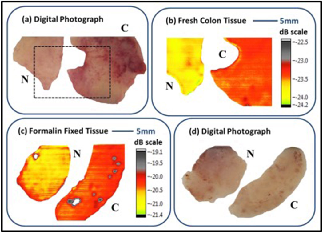
To investigate the suitability of continuous-wave THz imaging for detecting colorectal cancer, we acquired the reflectance from normal and cancerous regions and compared them. For this, we used a carbon dioxide (CO2) optically pumped far-IR gas laser (operating at 584GHz). We used a polarization-specific detection technique to obtain both co- and cross-polarized THz images of fresh and formalin-fixed colonic tissues and found that the reflection measurements exhibited an intrinsic contrast between normal and cancerous regions.1 Consequently, the cross- and co-polarized THz images exhibited increased reflection from the tumor. Our data analysis indicates that the cross-polarized THz images not only correlate better with the histology compared to the co-polarized ones, but also provide a constant relative-reflectance ratio (of normal versus cancerous tissue) that is independent of the patient.
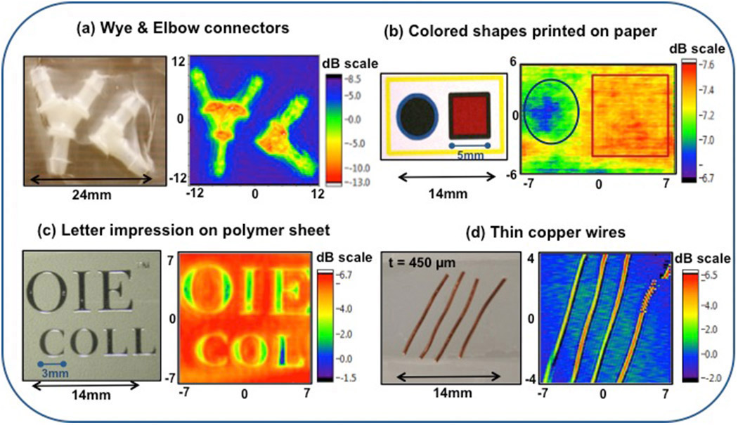
Finally, we built a prototype THz endoscopic system by integrating a thin metal-coated flexible THz waveguide with an imaging system. The THz source and detector are located remotely, such that neither is required to be inserted into the patient. As is illustrated in Figure 1, our imaging system uses a single channel to transmit and collect the back-reflected intrinsic THz signal from the sample, and is capable of operation in both transmission and reflection modalities.2 The THz images—see Figure 3—show that this approach has promise for applications in communication, sensing, and biomedical imaging. Additionally, the diameter of the THz waveguide is the only determining factor for the THz endoscope size. Because we are able to fabricate very small THz waveguides (with diameters ranging from a millimeter to hundreds of microns), THz endoscopes can be scaled down significantly according to practical requirements.6 The 2D images that we obtained using our prototype system show intrinsically higher THz reflectance from the tumor and adequately delineated abnormal regions—see Figure 4—thereby demonstrating the potential of THz imaging for cancer detection.7
 
Figure 3. Digital photographs (left) and THz images (right) of (a) nylon connectors, (b) solid shapes of different colors (blue and red) printed on white paper, (c) letters embossed on a polymer sheet, and (d) thin copper wires mounted on a glass slide. t: Thickness. dB: Decibel.
 
Figure 4. Digital photographs compared with cross-polarized THz reflection images of normal (N) versus cancerous (C) tissue: (a) and (b) show fresh colonic tissue sets; (c) and (b) show formalin-fixed colonic tissue sets. In both examples, the cancerous colonic tissue is clearly defined in the THz reflection images.
In summary, we have investigated the potential medical application of continuous-wave THz radiation for cancer detection and the feasibility of using a THz endoscope as an additional tool to aid in colorectal cancer screening. This proof-of-principle experiment demonstrates the possibility of obtaining an intrinsic endogenous contrast between normal and cancerous regions. The level of contrast that we attained represents a significant step toward the clinical endoscopic application of THz technology for real-time cancer screening. As a next step, we intend to test our prototype THz endoscopic system for in vivo measurements.

Pallavi Doradla
Wellman Center for Photomedicine
Harvard Medical School
Boston, MA
Pallavi Doradla received her PhD in physics from the University of Massachusetts Lowell and is now a postdoctoral researcher. Her research focuses on the fields of THz imaging and biomedical optics and her expertise lies in endoscopic imaging, catheter design, polarization-sensitive optical coherence tomography, flexible THz waveguide technology, and cancer detection.
Cecil Joseph, Robert H. Giles
Biomedical Terahertz Technology Center
University of Massachusetts Lowell
Lowell, MA

References:
1. P. Doradla, K. Alavi, C. Joseph, R. H. Giles, Detection of colon cancer by continuous-wave terahertz polarization imaging technique, J. Biomed. Opt. 18, p. 090504, 2013. doi:10.1117/1.JBO.18.9.090504
2. P. Doradla, Terahertz Endoscopic System for Cancer Detection, Lambert Academic Publishing, 2015.
3. C. S. Joseph, R. Patel, V. A. Neel, R. H. Giles, A. N. Yaroslavsky, Imaging of ex vivo nonmelanoma skin cancers in the optical and terahertz spectral regions optical and terahertz skin cancers imaging, J. Biophotonics 7, p. 295-303, 2014.
4. J. P. Martin, C. S. Joseph, R. H. Giles, Continuous-wave circular polarization terahertz imaging, J. Biomed. Opt. 21, p. 070502, 2016.
5. P. Doradla, R. H. Giles, Dual-frequency characterization of bending loss in hollow flexible terahertz waveguides, Proc. SPIE 8985, p. 898518, 2014. doi:10.1117/12.2038596
6. P. Doradla, C. S. Joseph, J. Kumar, R. H. Giles, Characterization of bending loss in hollow flexible terahertz waveguides, Opt. Express 20, p. 19176-19184, 2012. doi:10.1364/OE.20.019176
7. P. Doradla, K. Alavi, C. Joseph, R. H. Giles, Single-channel prototype terahertz endoscopic system, J. Biomed. Opt. 19, p. 080501, 2014. doi:10.1117/1.JBO.19.8.080501

Friday, November 18, 2011

Department of Defense awards $23 million dollar grant to use THz in surveillance radar imagery


In one of the largest grants awarded to the university in its history, a University of Massachusetts Lowell lab has received $23 million from the U.S. Army., in part thanks to Mansfield native Prof. Robert Giles.
The grant is for the UMass Submilimeter-Wave Technology Lab. The submilimeter-wave is term used to describe a type of rediation. Microwaves are bigger than, and infrared waves are smaller. The UMass lab primarily researches terahertz-frequency measurement systems, which have many different applications, from medical devices to military radar installations.
 The lab has developed and applied these technologies in the areas of military surveillance, homeland security, medical diagnostics and scientific and academic research.
The grant adds to 2006 funding from the U.S. Department of Defense, which awarded the lab $27 million to fund its research over five years.
“This latest grant is a continuation of our program to assist the government in acquiring and analyzing surveillance radar imagery,” Giles said. “It is a testament to and recognition of our high level of expertise in the field. Our research is focused on using terahertz-frequency sources and receivers to scale the Army’s millimeter-wave and microwave airborne radar systems.”
The research has been going on since 1979. From a military standpoint, the technology and research allowed for radar machines to get a defined and easily recognizable radar "footprint" for any type of aircraft or vehicle.  Such radar fingerprints are useful for quickly identifying whether an incoming object in the battlefield is a friend or foe.
“As a member of the Expert Radar Signature Solutions consortium developed by the National Ground Intelligence Center, we and our government sponsors are the only research program that uses terahertz-frequency measurement systems to collect real-world radar signature data,” Giles said.
On the medical side, they are useful for detecting non-melanoma skin cancer. Terahertz rays are non-ionizing and have no known harmful effects on living tissue. Also, terahertz rays have a shorter wavelength than microwaves, offering perhaps higher resolution for imaging applications.
Cecil Joseph, a post-doctoral researcher at the lab, has demonstrated there is sufficient contrast between healthy and cancerous tissue at terahertz frequencies. This could lead to a simpler and more cost-effective diagnostic tool for treating skin cancer.
“With sufficient external funding, we are hoping to build the hardware required for clinical studies,” Giles said.
In addition to its work for the Army, the research lab has used its unique capabilities to fulfill radar-measurement requests from other Department of Defense agencies as well as defense-related laboratories and companies, including MIT Lincoln Lab, Raytheon, Boeing and Lockheed-Martin.
For more information, go to http://stl.uml.edu.

Saturday, September 24, 2011

Research advances bring in vivo NMSC imaging closer to reality

Colors indicate increases or decreases in infr...Image via Wikipedia


Based on steady progress in laboratory studies, researchers are optimistic about the potential for noninvasive terahertz imaging systems to become a useful tool for intraoperative delineation of nonmelanoma skin cancers (NMSCs).
In a recent publication (Lasers Surg Med. 2011;43(6):457-462), Cecil S. Joseph, Ph.D., and colleagues from the Submillimeter-Wave Technology Laboratory, the Advanced Biophotonics Laboratory, at the University of Massachusetts, Lowell, and Massachusetts General Hospital, Boston, described positive results with continuous-wave terahertz transmission imaging for differentiating between cancerous and normal skin tissue.
At the 31st annual meeting of the American Society for Laser Medicine and Surgery, Dr. Joseph reported on the next step in development of the technology involving reflectance terahertz continuous-wave imaging.
“Terahertz imaging has several properties that make it attractive for in vivo imaging. At its present stage, our research with this technology is about one step removed from the development of a system that would be feasible for routine use in clinical practice,” Dr. Joseph says.
THz potential
H&E
An H&E histology of consecutive slice (far left), a 1.4 THz transmittance image (middle), and a 1.6 THz transmittance image, showing the cancerous area outlined by the black dotted line. (Photos: Cecil S. Joseph, Ph.D.)
Terahertz wavelengths lie between the microwave and infrared regions on the electromagnetic spectrum and are of interest for use in biomedical imaging because terahertz radiation has reasonable spatial resolution, is nonionizing, and a large number of biomolecules have characteristic resonance frequencies within the terahertz wavelength range.
For imaging of NMSCs, contrast with normal skin is intrinsic so that no exogenous contrast agent is needed and appears to be related to differences in water content and in the state of the water state (bound versus free) that exist between the malignant and normal tissue, Dr. Joseph says.
Wallace and Woodward, pioneers of terahertz imaging for NMSC, originally demonstrated its feasibility using a pulsed terahertz source, and they were able to show in in vivo experiments that it could be used to discriminate between basal cell carcinomas and noncancerous skin in reflection geometry.
Dr. Joseph and colleagues have been working to develop continuous-wave terahertz imaging, which would have a number of advantages compared with a pulsed system.
“A pulsed system typically needs a femtosecond laser to produce the desired frequency, whereas a continuous-wave system would likely be based on a solid-state source that would make it comparatively much less expensive to produce,” Dr. Joseph says.
“Furthermore, the signal-to-noise ratio is typically better with a continuous-wave source compared with a pulsed system, and the data-acquisition time is also faster because the data are collected at a single frequency,” he says.
Initial study
In an initial ex vivo study, Dr. Joseph and colleagues demonstrated transmission imaging of NMSC specimens using a continuous-wave terahertz imaging system. Using fresh tissue from Mohs surgeries performed at Massachusetts General Hospital, they first isolated the frequency that would produce contrast between cancerous and noncancerous regions, and then they identified the source receiver characteristics that would provide tumor demarcation.
The performance of the imaging system for cancer delineation was determined by correlating the terahertz images with hematoxylin and eosin histopathology.
A tool that could be used to identify cancer margins in vivo would have to work in reflection modality, however, so the researchers have gone on to generate reflection images with a continuous-wave terahertz imaging system. Now, they will be correlating those images with the transmittance images and histological findings, as well as evaluating the resolution.
“The next step will be to isolate specific parameters for frequency and amplitude stability that will be used to identify proper source technology. Once those specifications are determined, a clinically deployable source can be tested,” Dr. Joseph says.
“So far, we have used CO2 optically pumped far-infrared gas lasers as the terahertz source for our experiments,” he says. “These lasers have several advantages, as they have high power, are stable and allow for easy frequency selection. However, they are only suitable for use in a laboratory setting.
“Solid-state transceivers that can be used in clinical practice work with room temperature sources and detectors, but for adequate sensitivity, the detector has to have a very narrow frequency window to eliminate noise,” Dr. Joseph says.
“A priori knowledge of this target frequency will enable more efficient development of a continuous-wave terahertz reflection imaging system. The technology exists to build the system, but it is a matter of getting the parameters right,” he says.
Source: Modern Medicine.

Enhanced by Zemanta